

| Journal of Hematology, ISSN 1927-1212 print, 1927-1220 online, Open Access |
| Article copyright, the authors; Journal compilation copyright, J Hematol and Elmer Press Inc |
| Journal website http://www.thejh.org |
Letter to the Editor
Volume 4, Number 1, March 2015, pages 155-156
Gram Negative Bacteremia Diagnosed on Peripheral Blood Smear Examination
Chanchal Rana
Department of Pathology, Dr Lal Pathlabs, Lucknow, Uttar Pradesh 226006, India
Manuscript accepted for publication January 21, 2015
Short title: Bacteremia in PBS Examination
doi: http://dx.doi.org/10.14740/jh184w
| To the Editor | ▴Top |
Septicemia is a severe clinical syndrome characterized by systemic signs of infection, shock and systemic organ failure. Hence a rapid and definitive diagnosis is mandatory for proper management. Diagnosis of sepsis is routinely confirmed by presence of organisms in blood or at the site of infection which is usually done through culture which is a time consuming method and hence does not play much role in the initial treatment decision. In such scenario microscopic examination of peripheral blood can be of great help to hasten the confirmation of septicaemia, thereby enabling doctors to immediately select a more specific therapy [1-3]. Although several species of bacteria have been described on examination of peripheral blood smears but bacteremia due to gram negative rods has rarely been reported [1, 4-6].
We describe an interesting case of a 28-year-old female who presented with high grade fever which was associated with chills, vomiting, and loss of appetite and generalized weakness. Hematological examination revealed presence of anemia (hemoglobin 9.0 g/dL), leucocytosis (total leucocyte count 26.7 × 10³/μL) and thrombocytopenia (62 × 103/μL). Peripheral blood smear (PBS) examination showed neutrophilia (absolute neutrophil count of 21.89 × 10³/μL) and presence of shift leucocytes. Apart from these features, toxic vacuolizations were seen in the neutrophils along with frequent presence of intracellular structures which were uniform and rod shaped, suggestive of phagocytised bacilli. Similar bacilli were also seen lying extracellular in small and large clusters (Fig. 1). Gram stain of peripheral blood affirmed the presence of gram negative bacteremia. Later the blood culture confirmed the blood smear findings and revealed presence of Pseudomonas aureginosa. The patient’s condition kept on deteriorating and 2 days after admission, the female developed multi-organ failure and succumbed to her disease in spite of intensive medical treatment.
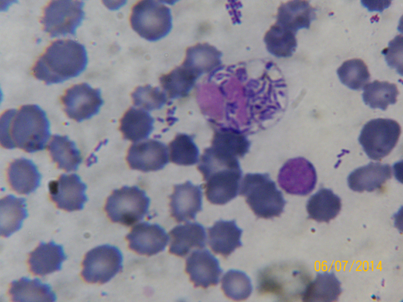 Click for large image | Figure 1. Leishman stained PBS (× 100; oil immersion) showing presence of intracellular (inside neutrophil) and extracellular rod shaped bacilli. Toxic vacuolizations are also seen inside the neutrophil. |
PBS has never been a popular diagnostic tool for septicemia due to certain limitations. Firstly, it is less sensitive technique as detection of microorganism requires a concentration of 105 CFU/mL or greater which is unusual [7, 8]. And secondly, it is the inability to identify the species of microorganism. For these reasons, this method is mainly limited for identification of parasitic organisms in diseases like malaria and filaria. Nevertheless this limitation can be overcome to certain extent. Sensitivity could be increased by using buffy coat along with increasing experience and sufficient training of laboratory observers [9]. Also typical gram reaction, morphologies and arrangements of the observed organisms may help in presumptive identification of certain etiological bacteria [10].
During PBS examination for bacteria certain points should be kept in mind. The organism should be intracellular and the cells containing them must be leucocytes. Associated finding like toxic granules, vacuoles and Dhole inclusion bodies in neutrophils are also very predictive of systemic infection. Presence of only extracellular microorganism should be reported with special care as it may represent contamination [11].
Thus to conclude, simple, inexpensive and safe PBS examination should be more widely used especially when overwhelming bacteremia is suspected in conditions like hyposplenism, AIDS neonatal sepsis, etc. In such scenario, it can prove to be an important tool to provide rapid preliminary diagnosis, thereby allowing clinicians to strengthen the empirical anti-microbial regimen.
Conflict of Interest
The authors declare that they have no conflict of interest.
| References | ▴Top |
- Fife A, Hill D, Barton C, Burden P. Gram negative septicaemia diagnosed on peripheral blood smear appearances. J Clin Pathol. 1994;47(1):82-84.
doi pubmed - Mirza I, Wolk J, Toth L, Rostenberg P, Kranwinkel R, Sieber SC. Waterhouse-Friderichsen syndrome secondary to Capnocytophaga canimorsus septicemia and demonstration of bacteremia by peripheral blood smear. Arch Pathol Lab Med. 2000;124(6):859-863.
pubmed - Sohn HE, Chung HR. Bacteremia diagnosed on peripheral blood smear before blood cultures becaome positive: a case report. Korean J Clin Pathol. 1999; 19:27-30.
- Takihi IY, Sandes AF. Killers on the road: Klebsiella and Pseudomonas bacteremia detected on peripheral blood smear. Blood. 2013;122(11):1851.
doi - Nakamura H, Saitou M, Kinjo S, Kaneshima H, Higa F, Tateyama M, Fujita J. Overwhelming pneumococcal bacteremia revealed by a peripheral blood smear in a 74-year-old healthy woman. Intern Med. 2007;46(6):303-306.
doi pubmed - Jeong TK, Jae HL, Hye SL, Yong GC, Dal SK, Sam IC, Soo CC. Bacteremia detected by a peripheral blood smear in a pediatrc surgical patient with thrombocytopenia. Korean J Clin Microbiol. 2010;13(4):182-185.
doi - Shanholtzer CJ, Schaper PJ, Peterson LR. Concentrated gram stain smears prepared with a cytospin centrifuge. J Clin Microbiol. 1982;16(6):1052-1056.
pubmed - Branda JA, Ferraro MJ, Kratz A. Sensitivity of peripheral blood smear review for the diagnosis of Candida fungemia. Arch Pathol Lab Med. 2007;131(1):97-101.
pubmed - Ristuccia PA, Hoeffner RA, Digamon-Beltran M, Cunha BA. Detection of bacteremia by buffy coat smears. Scand J Infect Dis. 1987;19(2):215-217.
doi pubmed - Misawa S. [Rapid diagnosis of infectious diseases; features and limitations of the microscopic examination of clinical specimens]. Rinsho Biseibutshu Jinsoku Shindan Kenkyukai Shi. 1999;10(2):121-131.
pubmed - Kroft SH. Infectious diseases manifested in the peripheral blood. Clin Lab Med. 2002;22(1):253-277.
doi
This is an open-access article distributed under the terms of the Creative Commons Attribution License, which permits unrestricted use, distribution, and reproduction in any medium, provided the original work is properly cited.
Journal of Hematology is published by Elmer Press Inc.