Journal of Hematology, ISSN 1927-1212 print, 1927-1220 online, Open Access
Article copyright, the authors; Journal compilation copyright, J Hematol and Elmer Press Inc
Journal website https://www.thejh.org

Case Report

Volume 11, Number 3, June 2022, pages 109-112


Non-Hepatosplenic Extramedullary Hematopoiesis Masquerading as Metastatic Malignant Melanoma

Figures

Figure 1.
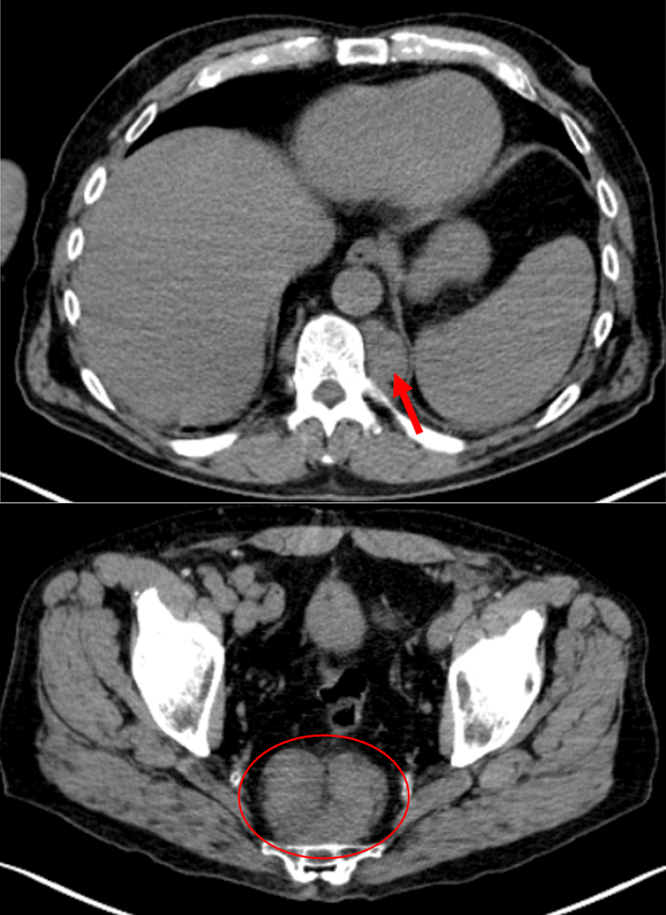
Figure 1. PET-CT images revealing a paraspinal mass at the level of T11 measuring 3.7 × 2.4 cm with SUV 2.7 (arrow) and the biopsy site presacral nodule measuring 7.5 × 5 cm with SUV 3.0 (circle). PET-CT: positron emission tomography-computed tomography; SUV: standardized uptake value.
Figure 2.
Figure 2. (a) Biopsy of presacral mass demonstrates NHS-EMH with trilineage hematopoietic cells including megakaryocytes (black arrows), myelocytes (blue arrows) and erythrocytes (red circles). (b) Blood vessel identified with surrounding areas trilineage hematopoiesis within the presacral mass (arrow). NHS-EMH: non-hepatosplenic extramedullary hematopoiesis.