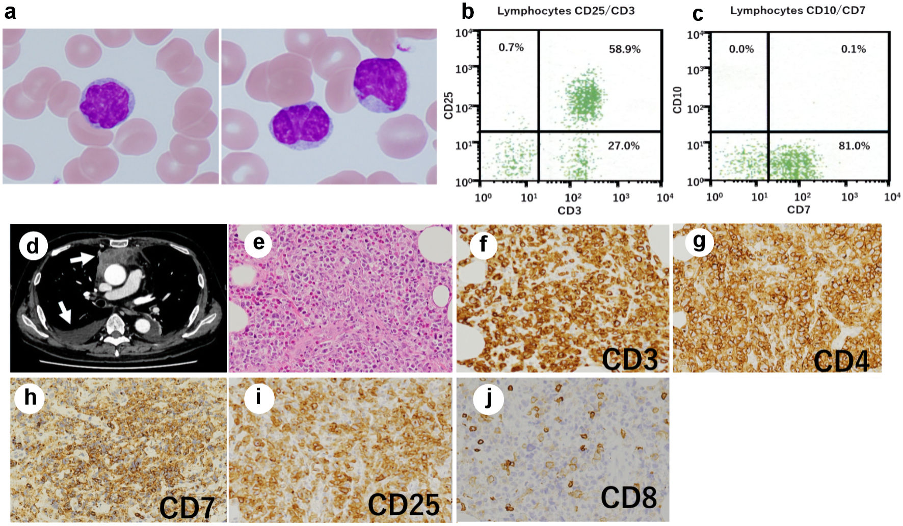
Figure 1. Posttransplant expansion of atypical T cells in the recipient. (a) Atypical lymphocytes with nuclear lobulation were observed in the recipient’s peripheral blood after transplantation; original magnification × 1,000, Wright-Giemsa stain. (b, c) Flow cytometry revealed T cells expressing CD3 (b), CD25 (b), and CD7 (c). (d) Computed tomography of the chest showed a mediastinal tumor and pleural effusion (arrows). (e-j) Biopsy of the mediastinal tumor was performed, showing an infiltrate of medium- to large-sized atypical lymphocytes (original magnification × 200). (e) Hematoxylin and eosin stain. The atypical cells were positive for CD3 (f), CD4 (g), CD7 (h), and CD25 (i), and negative for CD8 (j).
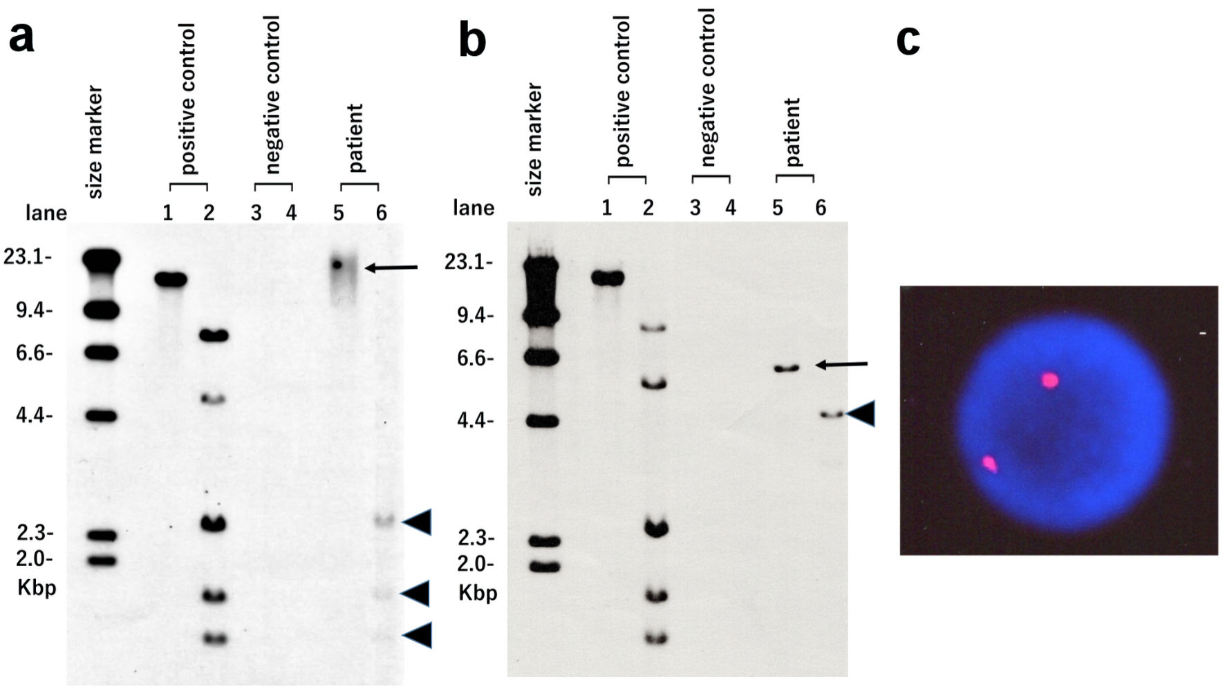
Figure 2. HTLV-I integration analysis using Southern blotting and XY-fluorescence in situ hybridization (XY-FISH). Atypical lymphocytes observed in the recipient after transplantation (a) and pretransplant ATLL cells of the recipient (b) were used. Lane 1: positive control in EcoR1 digestion. Lane 2: positive control in Pst1 digestion. Lane 3: negative control in EcoR1 digestion. Lane 4: negative control in Pst1 digestion. Lane 5: patient sample in EcoR1 digestion (arrows). Lane 6: patient sample in Pst1 digestion (arrowheads). (c) XY-FISH (X chromosome red and Y chromosome green) of atypical lymphocytes observed in the recipient after transplantation was performed. No XY signal was found in the nuclei of atypical lymphocytes. HTLV-I: human T-lymphotropic virus type I.
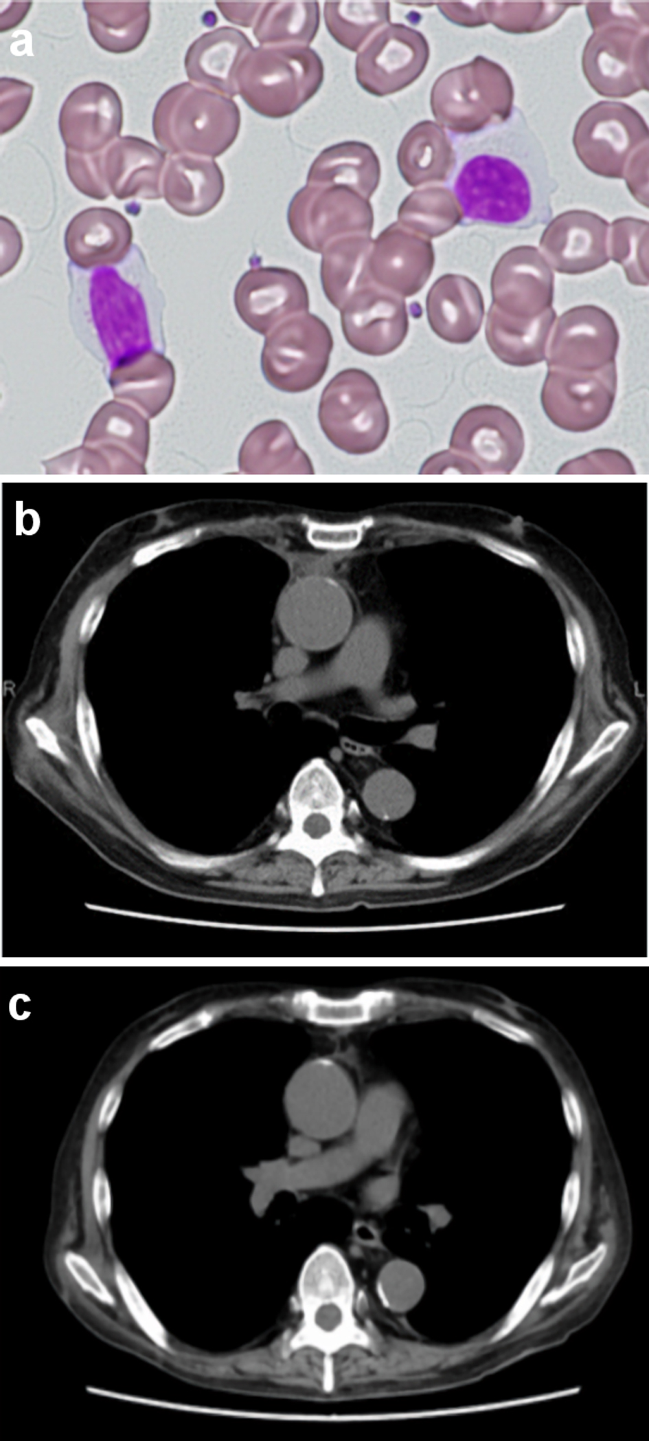
Figure 3. Peripheral blood lymphocytes and the mediastinal tumor after discontinuation of tacrolimus. No atypical lymphocytes with nuclear lobulation were observed in the recipient’s peripheral blood 2 months after discontinuation of tacrolimus (a) Original magnification × 400, Wright-Giemsa stain. (b, c) Computed tomography of the chest showed that the mediastinal tumor and pleural effusion regressed and then disappeared following discontinuation of tacrolimus ((b) Two months after discontinuation of tacrolimus. (c) Four months after discontinuation of tacrolimus).