Journal of Hematology, ISSN 1927-1212 print, 1927-1220 online, Open Access
Article copyright, the authors; Journal compilation copyright, J Hematol and Elmer Press Inc
Journal website https://www.thejh.org

Original Article

Volume 11, Number 1, February 2022, pages 8-14


Hematopoietic Stem Cell Transplantation Stabilizes Cerebral Vasculopathy in High-Risk Pediatric Sickle Cell Disease Patients: Evidence From a Referral Transplant Center

Figure

Figure 1.
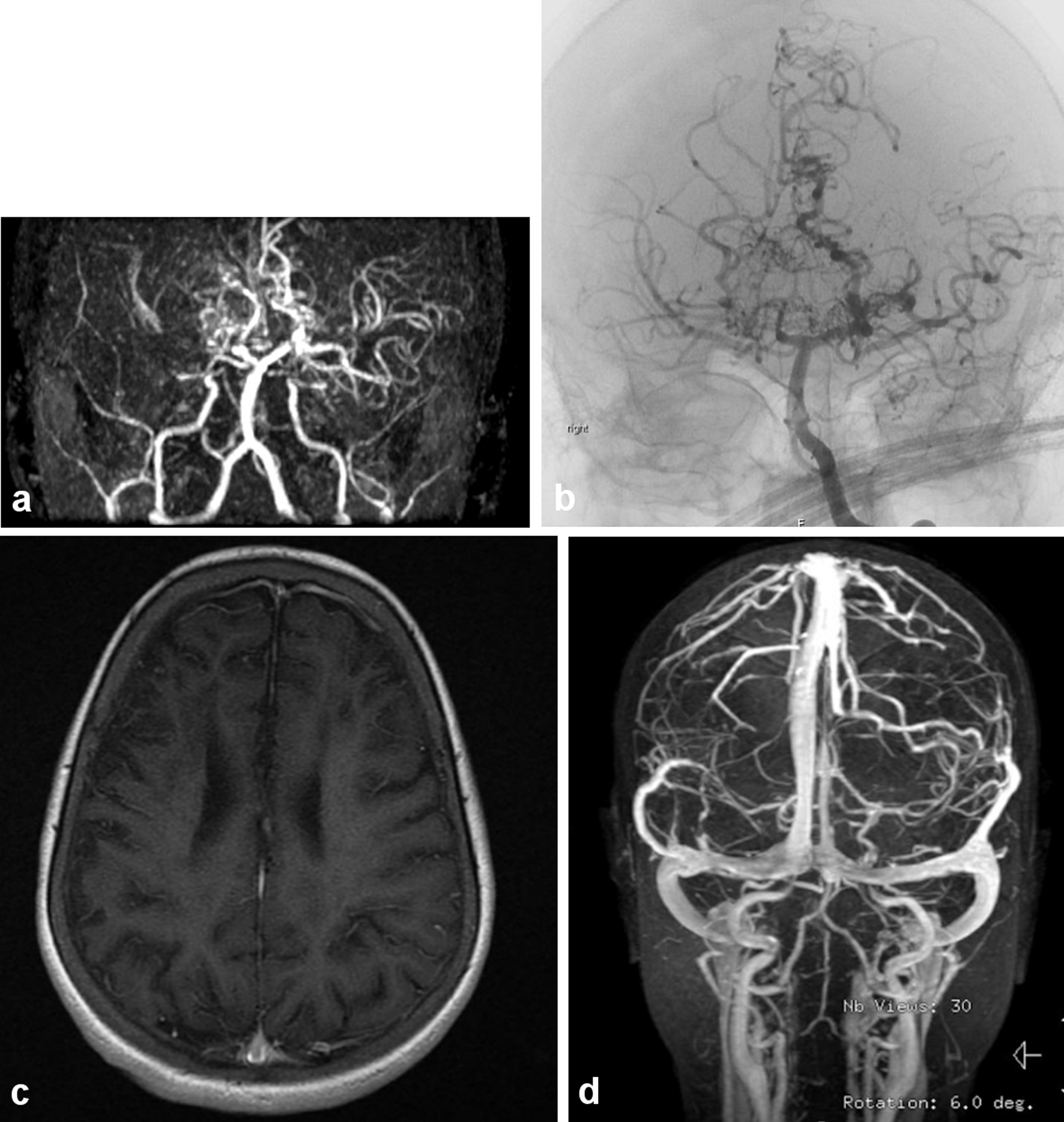
Figure 1. (a) The magnetic resonance (MR) angiography of the intracranial circulation showed significant prominent collateral vessels in the posterior cerebral artery territory with evidence for probable retrograde filling of middle cerebral artery territory from the posterior circulation. Significant collateral vessel prominence especially in the perforator branches in the thalamic region is detected. The findings are consistent with moyamoya disease with major thinning and attenuation in the supraclinoid internal carotid artery, and associated multiple watershed zone infarctions (before HSCT: March, 2009). (b) Evidence of narrowing at the distal internal carotid artery in the supraophthalmic segment bilaterally with the development of significant amount of small perforators giving puff of smoke appearance characteristic of moyamoya disease (same patient after HSCT in July, 2011). (c) Significant cortical and subcortical encephalomalacia diffusely along supratentorial brain parenchyma, significant within right frontal lobe with evidence of hemodynamic ischemic changes along the bilateral centrum semiovale. (d) Magnetic resonance imaging (MRI) with normal vascular distribution with moderately severe sickle cell disease.

Tables

Table 1. Patient Characteristics and Primary Disease-Related Parameters
 
Values are provided as numbers (%) unless otherwise specified. HSCT: hematopoietic stem cell transplant.
Age at infusion, median (range), years10.6 (3.0 - 13.9)
Gender
  Female20 (80.0%)
  Male5 (20.0%)
Sickle cell disease
  SS22 (88.0%)
  Sβ03 (12.0%)
Indications for HSCT
  Cerebrovascular accident (n = 25)
    Severe with moyamoya changes14 (56.0%)
    Moderate infarction7 (28.0%)
    Silent2 (8.0%)
    Transient2 (8.0%)
Symptoms (symptomatic = 23, 92%)
  Convulsions13 (52.0%)
  Hemiparesis11 (44.0%)
  Headache6 (24.0%)
  Psychiatric symptoms5 (20.0%)
  Focal neurological loss4 (16.0%)
  Loss of consciousness (any episode)2 (8.0%)


Table 2. Post-Transplant Primary Disease Evaluation
 
Outcome of interestPre-HSCT, n (%)Post-HSCT, n (%)Improvement, n (%)P value
aThe patient died after 6.5 months post-infusion due to exacerbation of chronic GVHD of gut. HSCT: hematopoietic stem cell transplant; GVHD: graft-versus-host disease; CV: cerebral vasculopathy.
Clinical findings pre- and post-HSCT
  Hemiparesis11 (44.0%)1 (4.0%)10 (90.9%)0.002
  Headache6 (24.0%)1 (4.0%)5 (83.3%)0.063
  Psychological symptoms5 (20.0%)2 (8.0%)3 (60.0%)0.250
  Focal neurological loss4 (16.0%)2 (8.0%)2 (50.0%)0.500
  Loss of consciousness (any episode)2 (8.0%)1 (4.0%)1 (50.0%)1.000
  Convulsions13 (52.0%)8 (32.0%)5 (38.5%)0.063
Post-HSCT radiological imaging results (n = 15)
  Stabilized CV (alive)14 (93.3%)
  Stabilized CV (expired)a1 (6.1%)