Journal of Hematology, ISSN 1927-1212 print, 1927-1220 online, Open Access
Article copyright, the authors; Journal compilation copyright, J Hematol and Elmer Press Inc
Journal website https://www.thejh.org

Case Report

Volume 10, Number 5, October 2021, pages 217-220


Acute Respiratory Distress Syndrome in a Patient With Acute Promyelocytic Leukemia: Overlapping Between Differentiation Syndrome and COVID-19

Figures

Figure 1.
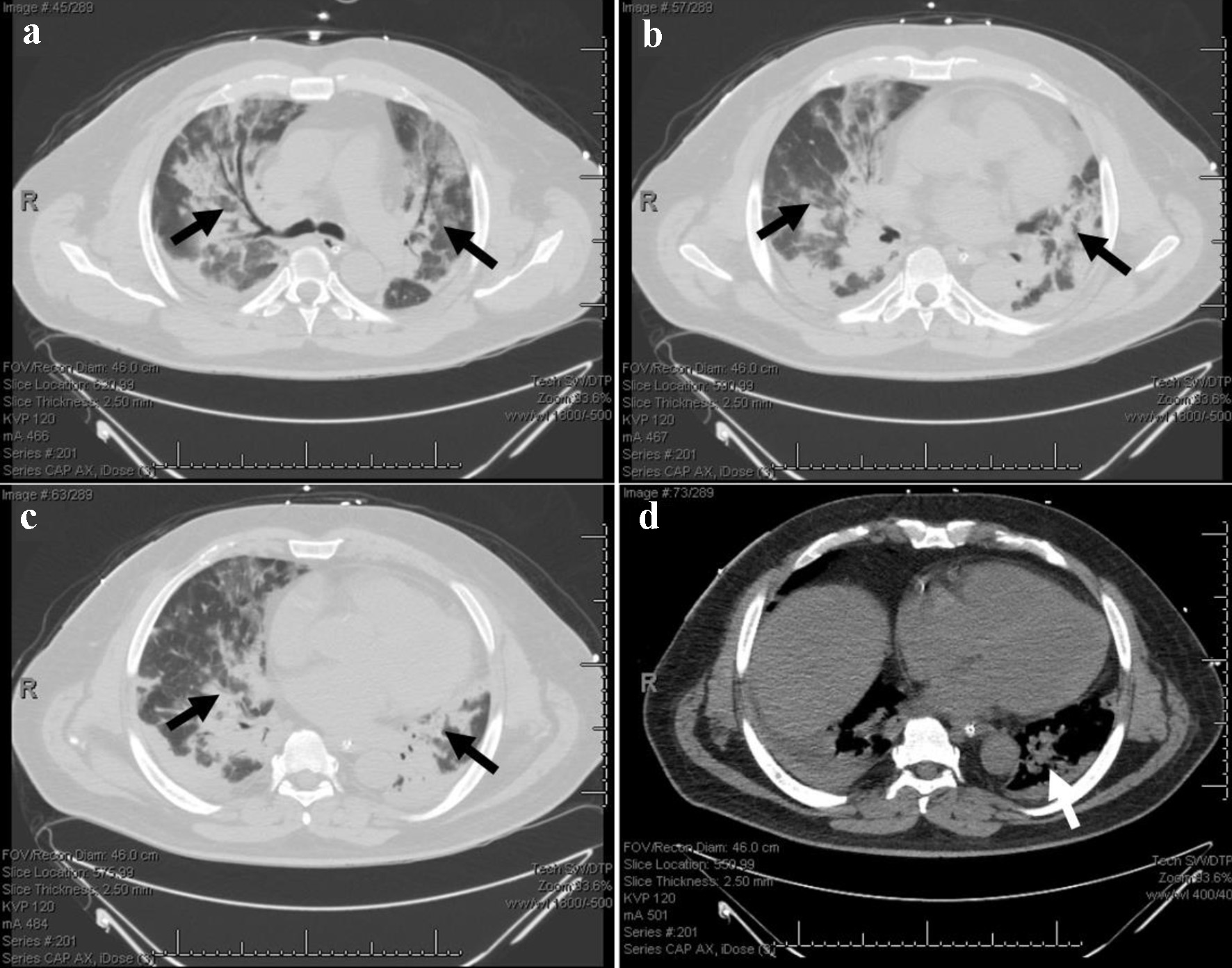
Figure 1. Chest CT on day 6 after admission. (a-c) Bilateral ground-glass opacities and superimposed consolidations. (d) Trace bilateral pleural effusions, right greater than left. The arrows indicate the lesions. CT: computed tomography.
Figure 2.
Figure 2. Continuous monitoring of complete blood count, creatinine, and coagulation during hospitalization. (a) White blood cell count (reference range, 4.0 - 11.0 × 10⁹/L). (b) Red blood cell count (reference range, 4.40 - 5.80 × 10¹²/L). (c) Platelet count (reference range, 150.0 - 400.0 × 10⁹/L). (d) Creatinine (reference range, 0.7 - 1.3 mg/dL). (e-h) Coagulation testing, including PT (reference range, 12.0 - 14.5 s), aPTT (reference range, 25.0 - 35.0 s), fibrinogen (reference range, 220.0 - 498.0 mg/dL), and D-dimer (reference range, 0.0 - 240.0 ng/mL). PT: prothrombin time; aPTT: activated partial thromboplastin time.
Figure 3.
Figure 3. A brief summary of patient management during hospitalization. DIC: disseminated intravascular coagulation; DS: differentiation syndrome; ECMO: extracorporeal membrane oxygenation.