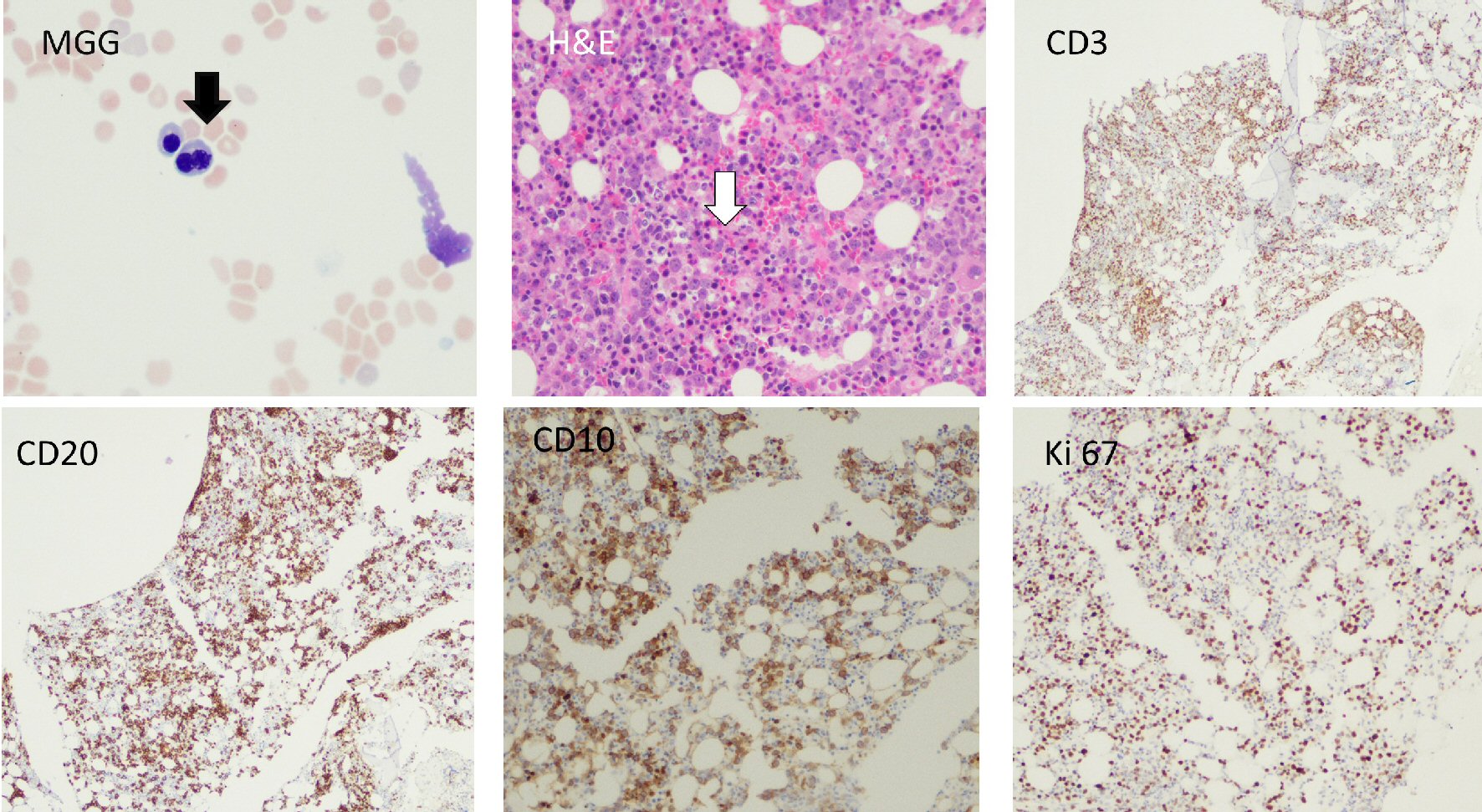
Figure 1. Bone marrow aspirate, trephine biopsy and immunohistochemical stains are shown. Black arrow shows a bilobed normoblast. White arrow shows presence of abnormal localization of immature precursors (ALIP). CD10, CD20 and Ki67 stains show infiltration of clonal large B-lymphocytes with a moderately high proliferative index (Ki67, 40-50%). H&E: hematoxylin and eosin.
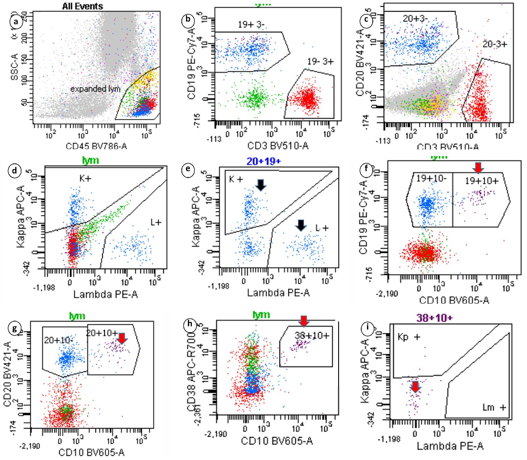
Figure 2. Sequential gating strategy identified the lymphocytes in the side scatter versus CD45 plot. The isolated lymphocytes (20+19+ gates) showed polytypic expression of surface immunoglobulins (black arrow). Events that expressed CD19, CD10 and CD38 highlighted the hematogone population, which lacked surface immunoglobulins (red arrows). Clonal B-lymphocytes seen on immunohistochemistry were not captured.