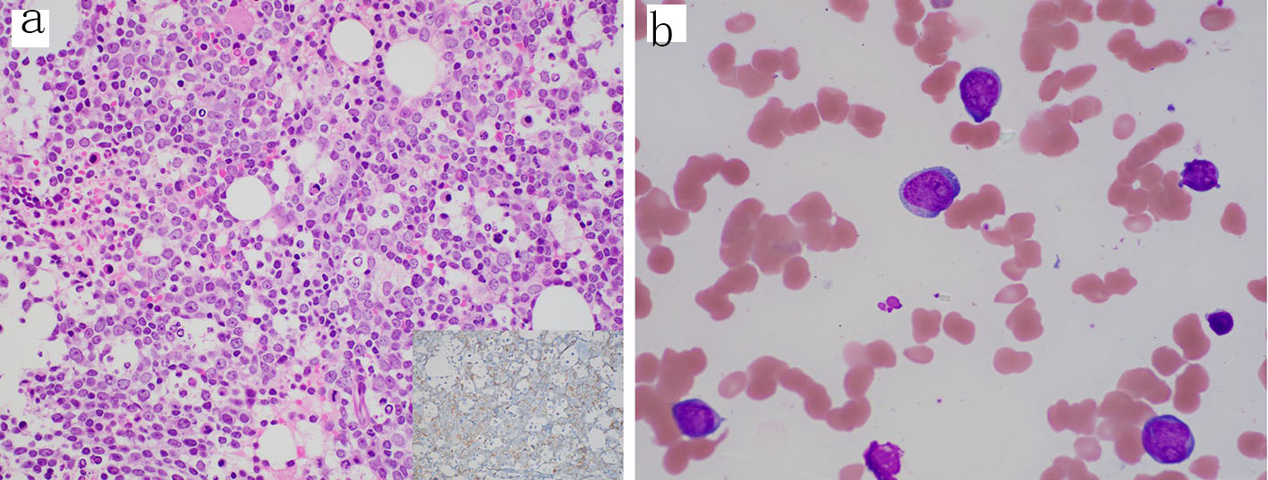
Figure 1. Diagnostic bone marrow biopsy and aspirate with acute myeloid leukemia. (a) Core biopsy showing sheets of blasts and decreased trilineage hematopoiesis (H&E, 400 ×); inset, CD34 stain (400 ×) highlighting the increased blasts. (b) Aspirate smear showing blasts with irregular nuclei, fine chromatin, prominent nucleoli and scant cytoplasm (Wright-Giemsa, 1000 × oil immersion). H&E: hematoxylin-eosin staining.
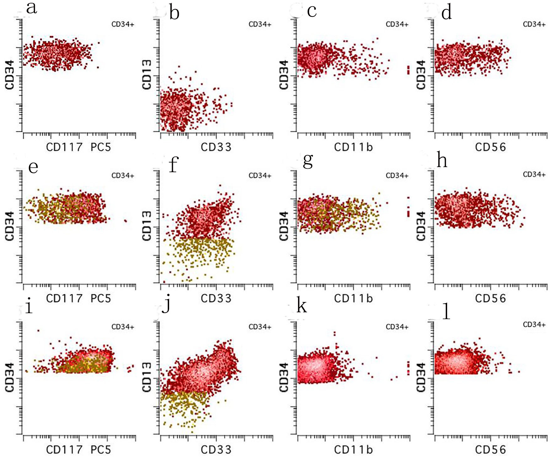
Figure 2. Ten color multiparameter flow cytometry. (a-d) Flow cytometry performed on the peripheral blood at our institution shortly after (day 3) the diagnostic bone marrow was taken showing a CD34 positive blast population (3.7% of total white blood cells) with abnormal expression of CD117 (absent to dim), CD13 (absent), CD33 (mostly absent), and CD56 (subset). (e-h) Flow cytometry performed on the day 21 bone marrow aspirate showing a small abnormal blast population (orange, 0.08% of total white blood cells) with a similar immunophenotype to those seen in the peripheral blood on day 3. (i-l) Flow cytometry performed on the day 49 bone marrow aspirate showing no abnormal myeloid blast population.
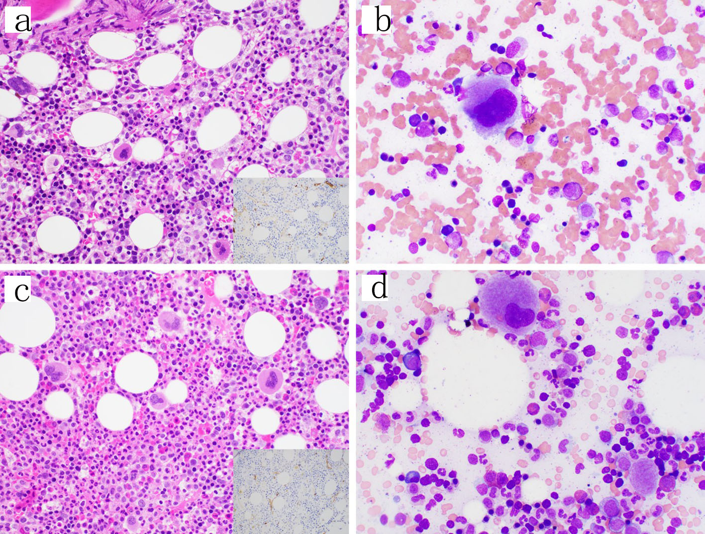
Figure 3. Follow-up bone marrow biopsies and aspirates. (a, b) Day 21 from the original bone marrow biopsy. The core biopsy showed trilineage maturing hematopoiesis and no increase in blasts by H&E (400 ×) or CD34 immunohistochemical stain (inset, 400 ×) or the aspirate smear (1000 × oil immersion); however, a small abnormal blast population was detected by flow cytometry. (c, d) Day 49 from the original bone marrow biopsy similarly showing trilineage maturing hematopoiesis and no increase in blasts on the core biopsy by H&E (400 ×) or CD34 immunohistochemical stain (inset, 400 ×) or the aspirate smear (1000 × oil immersion) and no abnormal blast population detected by flow cytometry. H&E: hematoxylin-eosin staining.