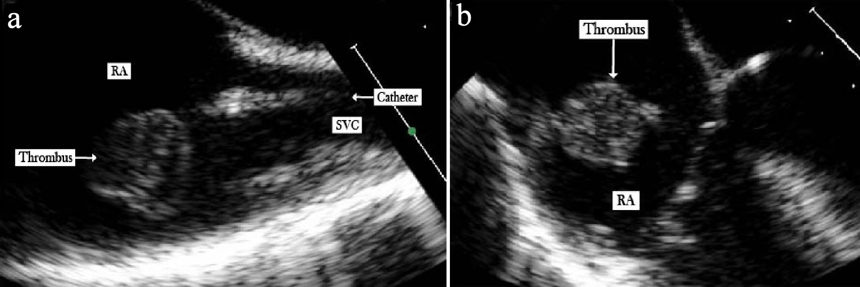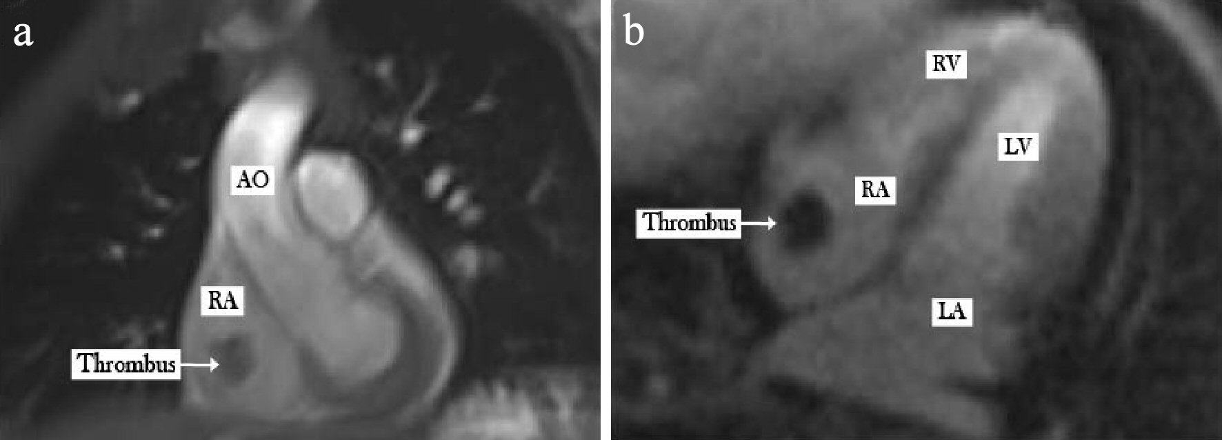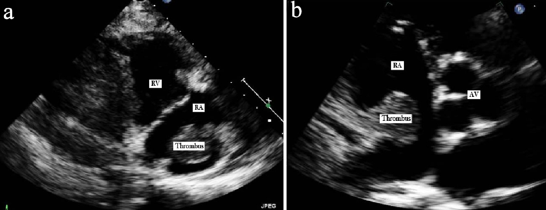
Figure 1. Transthoracic echocardiogram. (a) Parasternal right ventricle inflow view illustrating the RA thrombus. (b) Short axis view showing the RA thrombus attached to the posterior wall through a stalk.
| Journal of Hematology, ISSN 1927-1212 print, 1927-1220 online, Open Access |
| Article copyright, the authors; Journal compilation copyright, J Hematol and Elmer Press Inc |
| Journal website http://www.thejh.org |
Case Report
Volume 8, Number 3, September 2019, pages 125-128
Incidental Finding of Right Atrial Mass in a Patient With Hodgkin Lymphoma: Utilization of Multimodal Cardiac Imaging in Diagnosis
Figures