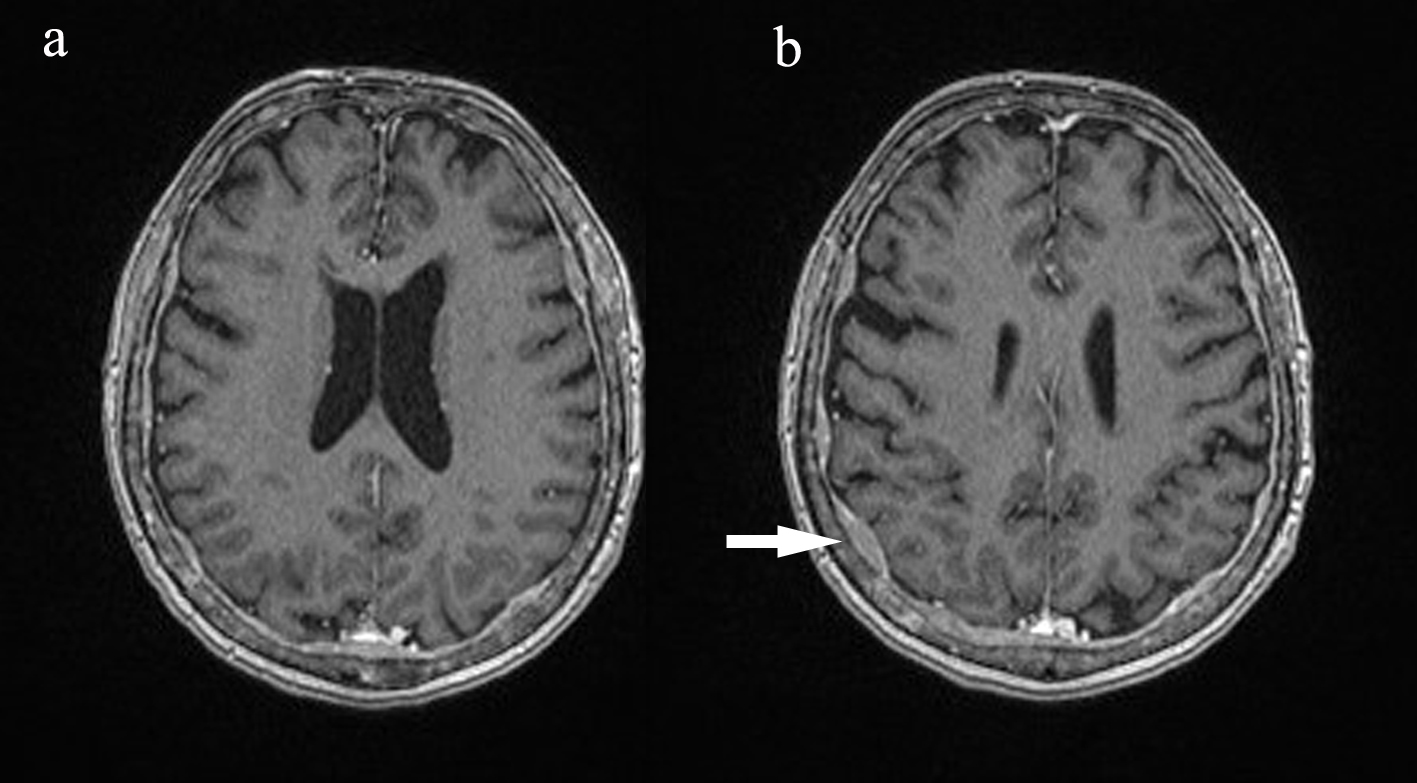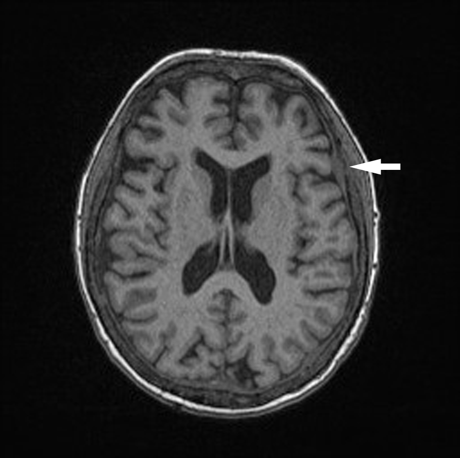
Figure 1. Unenhanced axial T1 MPR brain. Dural thickening diffusely.
| Journal of Hematology, ISSN 1927-1212 print, 1927-1220 online, Open Access |
| Article copyright, the authors; Journal compilation copyright, J Hematol and Elmer Press Inc |
| Journal website http://www.thejh.org |
Case Report
Volume 8, Number 1, March 2019, pages 29-33
Intracranial Involvement in Multiple Myeloma Presenting as a Cranial Nerve Palsy
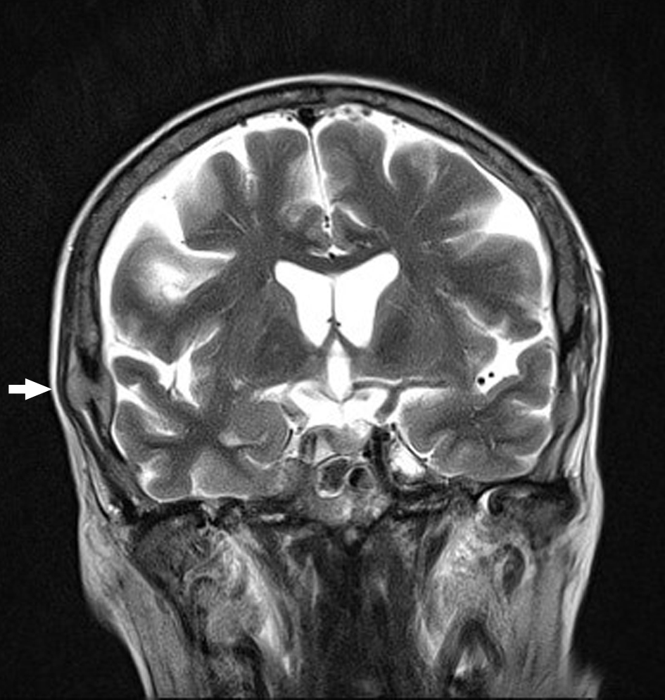
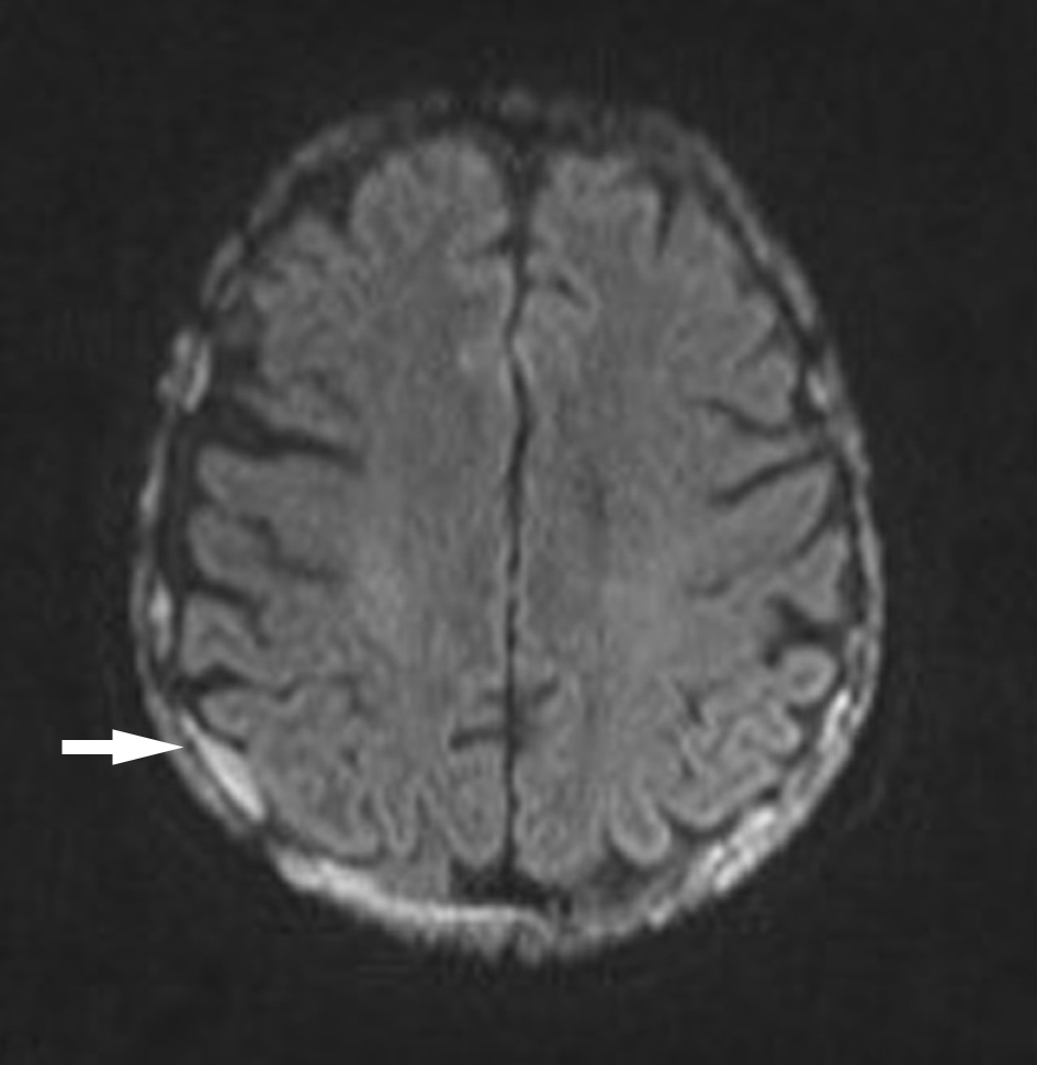
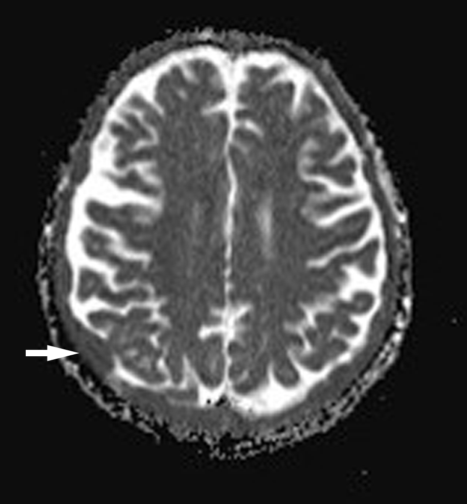
Figures