Figures
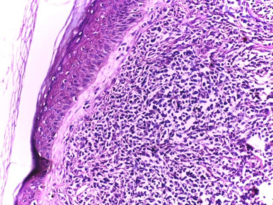
Figure 1. Infiltration of the dermis and hypodermis by monomorphous non-epidermotropic diffuse infiltrate of small to medium-sized cells with irregular nuclear contours. The cellular infiltrate is separated from the epidermis by a Grenz zone (H&E, × 200).
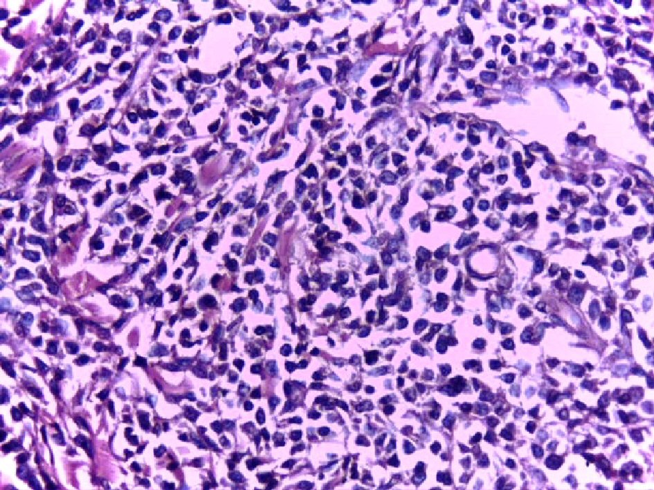
Figure 2. High power of the same case showed infiltrate of small to medium-sized cells with irregular nuclear contours, pleomorphic nuclei and scant amount of cytoplasm (H&E, × 200).
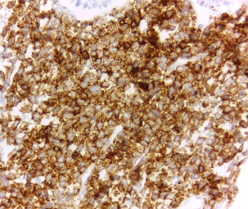
Figure 3. Strong membranous staining of the tumor cells for CD45 (DAB, × 400).
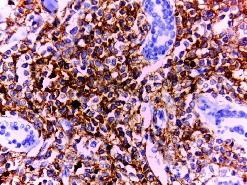
Figure 4. Diffuse positive membranous staining of the tumor cells for CD4 (DAB, × 400). Note periadnexal aggregation of the tumor cells.
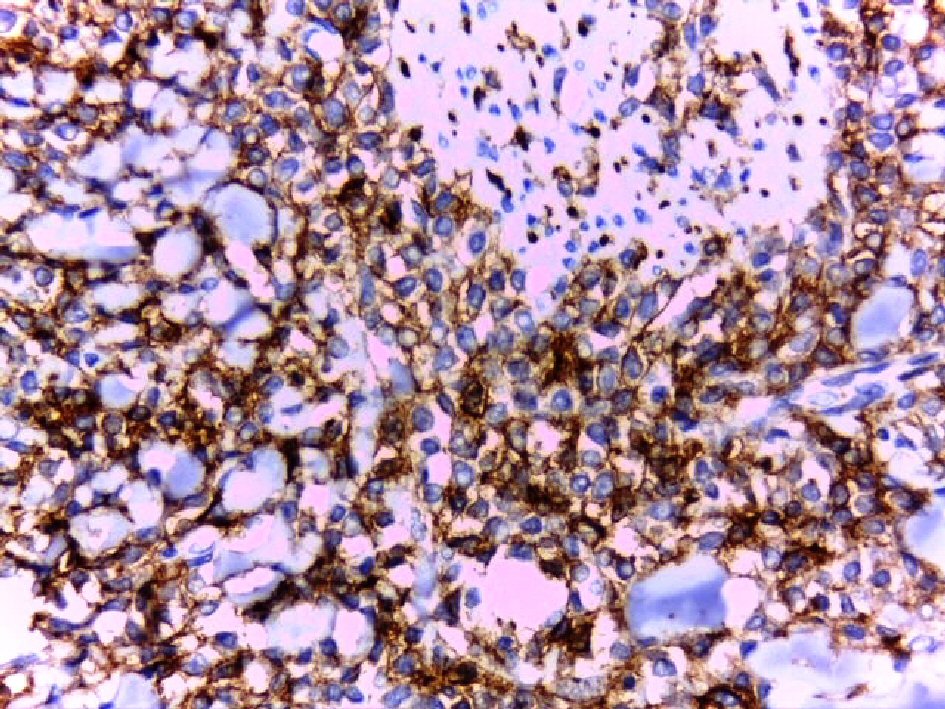
Figure 5. Strong membranous staining of the tumor cells for CD 56 (DAB, × 400).
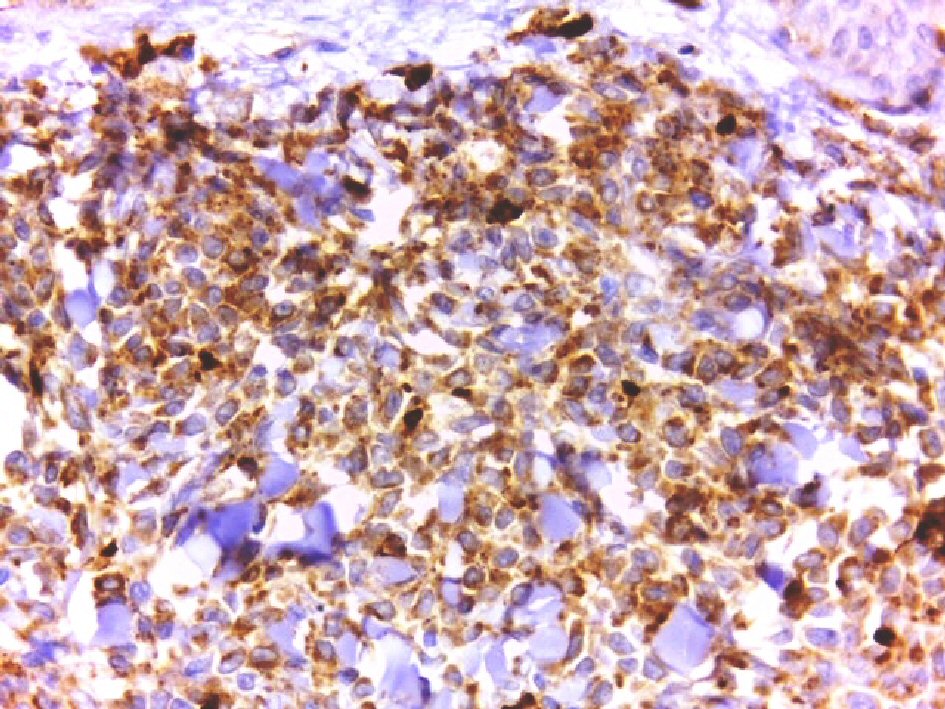
Figure 6. Positive cytoplasmic staining of the tumor cells for CD 68 (DAB, × 400).