Figures
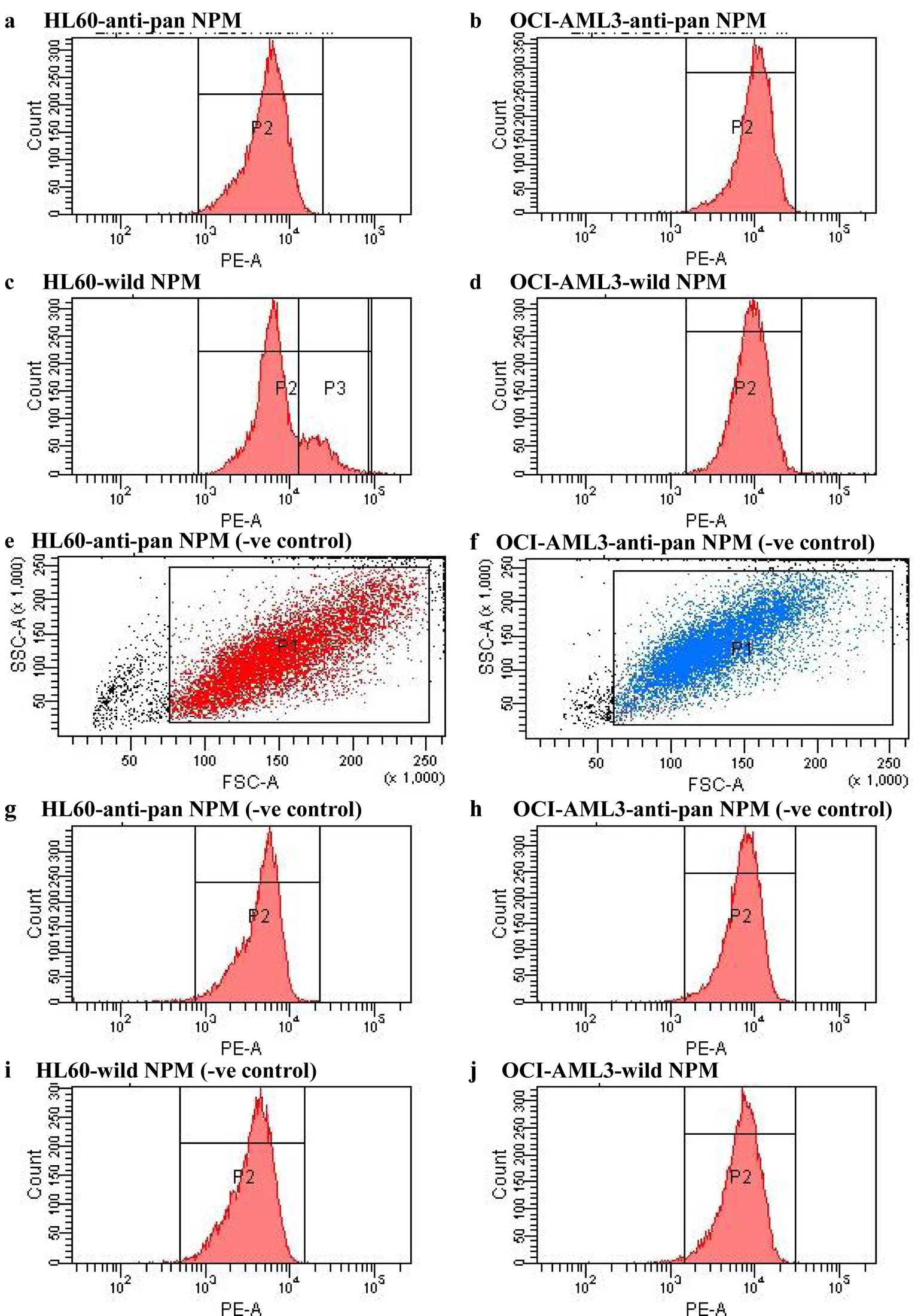
Figure 1. FACS analysis of HL60 and OCI-AML3 cells labeled with specific anti-NPM antibodies including anti-pan NPM and antibody for wild-type NPM, and their equivalent negative controls as labeled in the figure. NPM: nucleophosmin; FACS: fluorescence-activated cell sorting; AML: acute myeloid leukemia.
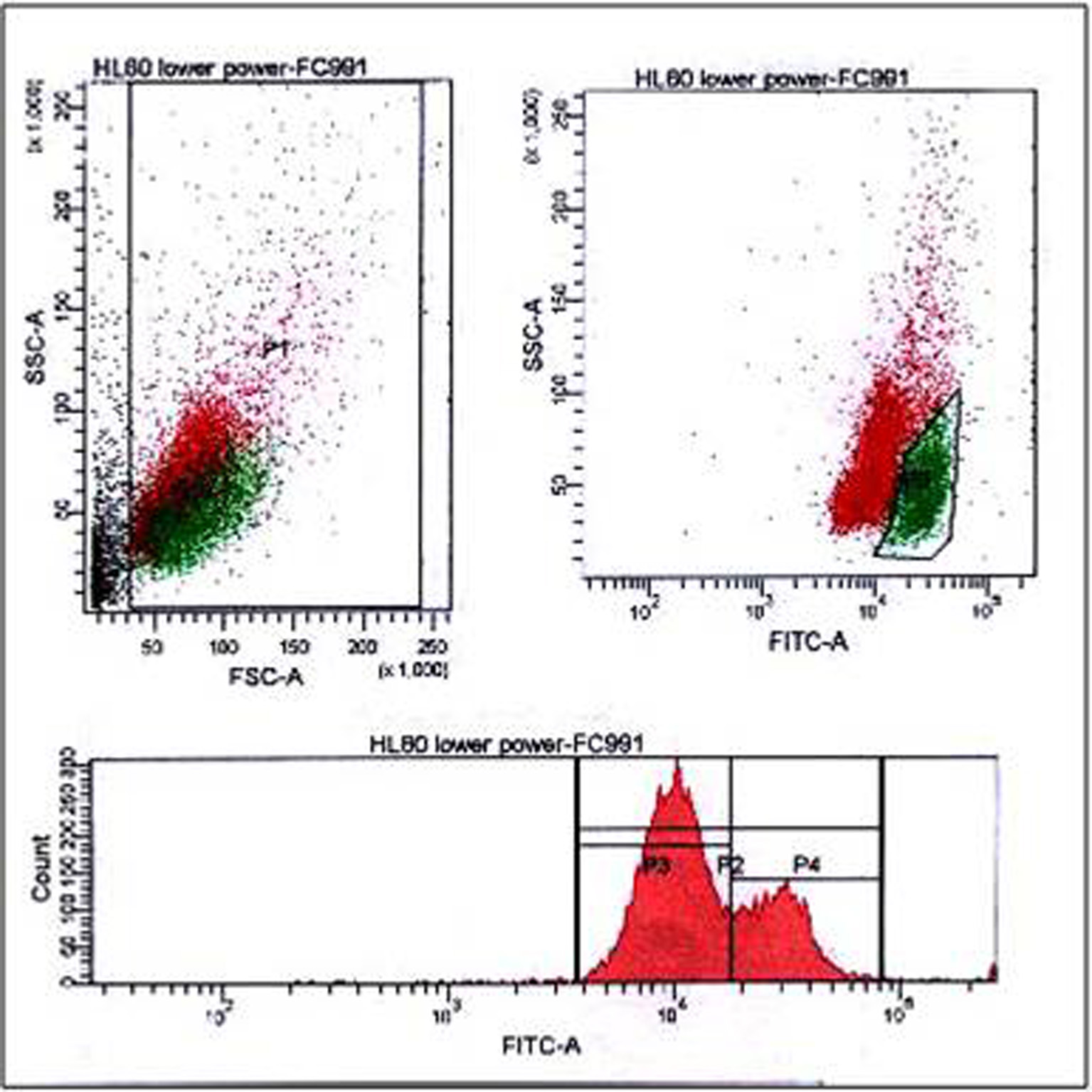
Figure 2. FACS analysis of HL60 cells labeled with wtNPM, showing dual populations in the bivariate dot plots (upper panel), quiescent (red) and dividing (green), both cellular debris and dividing cells were gated as indicated in the figure, while the lower panel showing a humped peak single parameter histogram. wtNPM: wild-type nucleophosmin; FACS: fluorescence-activated cell sorting.
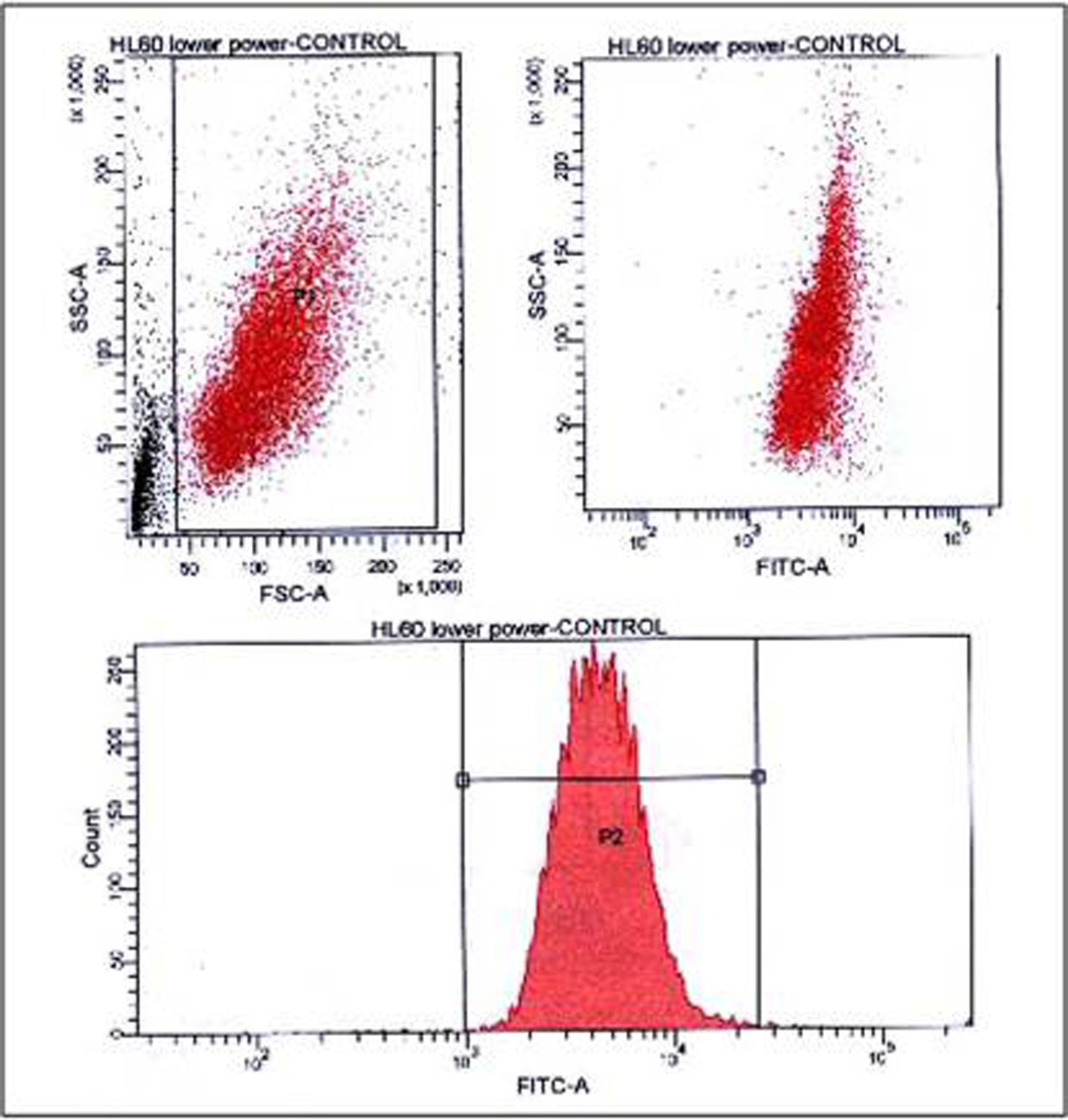
Figure 3. FACS analysis of HL60 negative control (secondary antibody only), showing presence of single population in the bivariate dot plots (upper panel), debris was gated as indicated, and a single peak MF histogram in the lower panel. FACS: fluorescence-activated cell sorting.
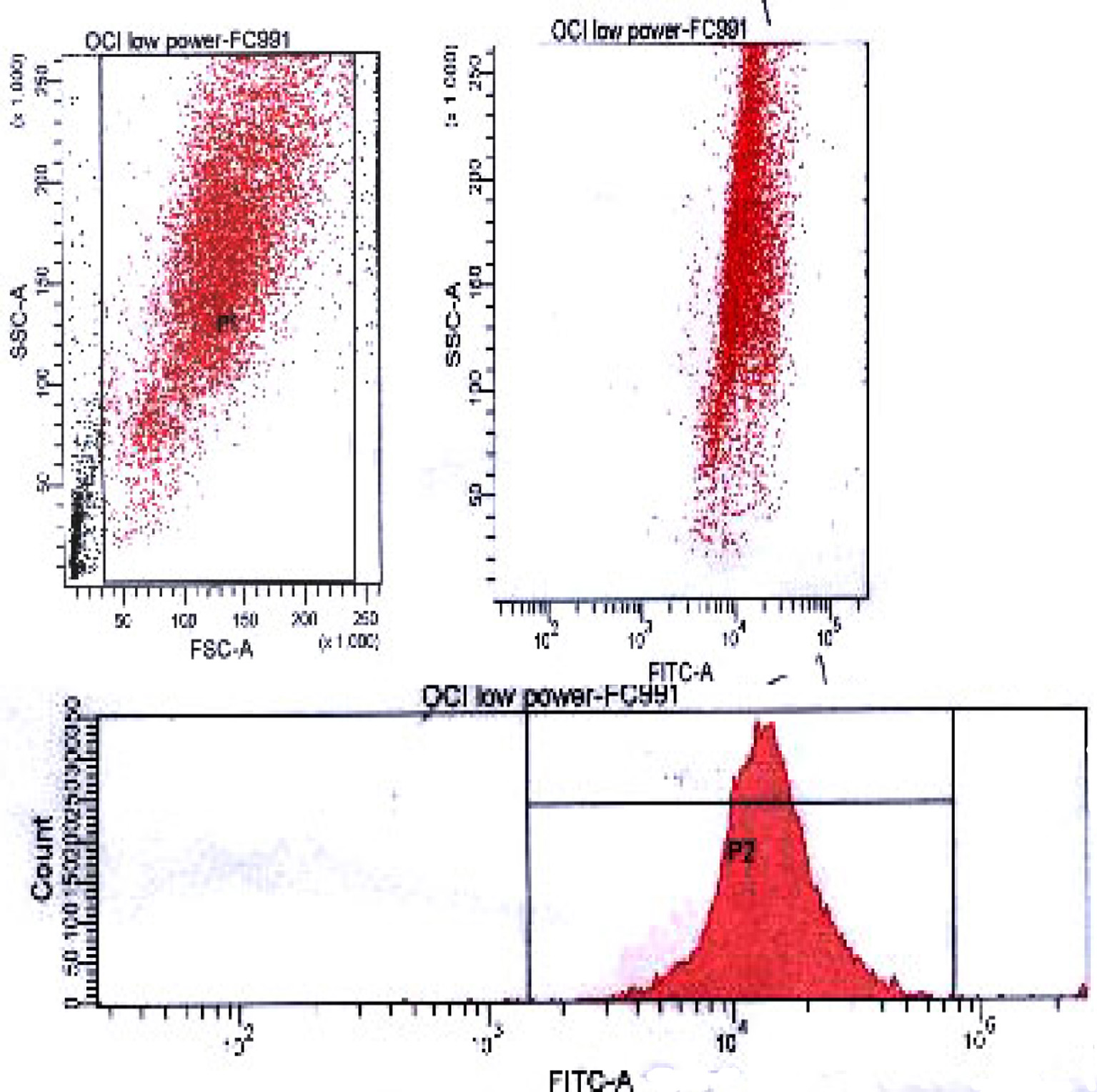
Figure 4. FACS analysis of wtNPM in OCI-AML3 cells, showing a single population in the bivariate dot plots (upper panel), debris was gated as indicated, and a single peak MF histogram in the lower panel. FACS: fluorescence-activated cell sorting; wtNPM: wild-type nucleophosmin; AML: acute myeloid leukemia.
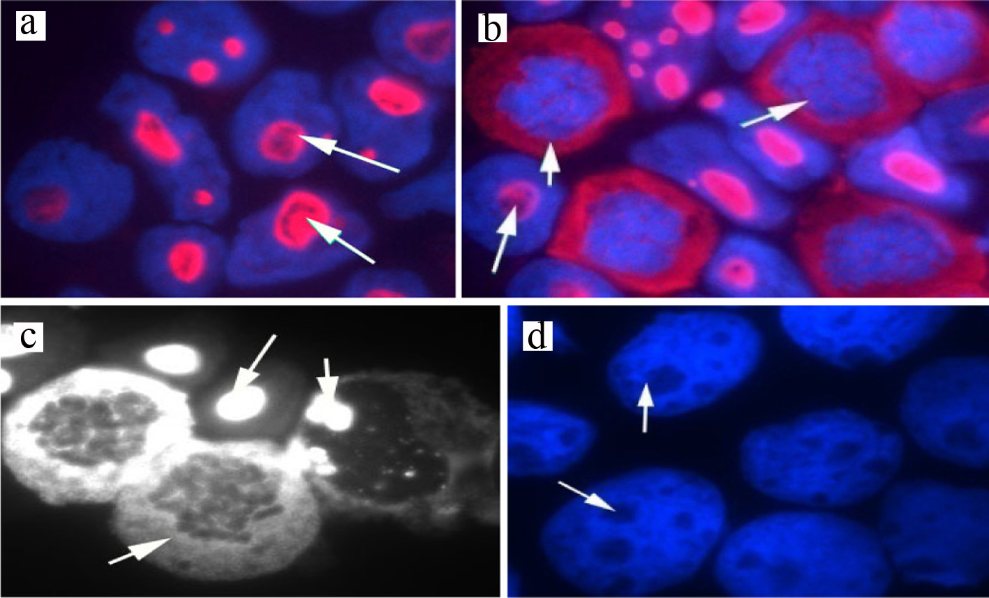
Figure 5. Immunostaining of wtNPM in (a) resting HL60 showing nucleolar localization (red arrowed), (b) dividing HL60 showing cytoplasmic localization in the arrowed cells, images were captured with the Hamamatasu camera and the nuclei were stained with DAPI (blue), (c) HL60 cells stained with wtNPM (white) image, grabbed with the Nikon Eclipse black and white camera, (d) HL60 negative control stained with Texas Red-conjugated secondary antibody only (no staining detected). wtNPM: wild-type nucleophosmin; DAPI: 4',6-diamidino-2-phenylindole.
Tables
Table 1. Numerical Values of Flow Cytometeric Analysis of Human Leukemia Cell Lines, HL60 and OCI-AML3 Labeled With wtNPM and Anti-Pan NPM, and Their Equivalent Negative Controls
| Test/control | % patients | SSC (mean) | PE (mean) |
|---|
| wtNPM: wild-type nucleophosmin; SSC: side scatter; PE: phycoerythrin; AML: acute myeloid leukemia. |
| OCI-AML3 anti-pan NPM | 99.9 | 131,484 | 10,820 |
| HL60-anti-pan NPM | 99.6 | 129.079 | 5,779 |
| OCI-AML3 wtNPM | 99.0 | 125,566 | 9,855 |
| HL60 wtNPM |
| P1 | 98.7 | 120,507 | 9,522 |
| P2 | 18.4 | 101,113 | 23,654 |
| HL60 control wt | 98.4 | 120,468 | 4,900 |
| OCI-AML3 control wt | 99.8 | 131,925 | 7,748 |
| OCI-AML3 control anti-pan | 99.7 | 132,628 | 7,509 |
| HL60 control anti-pan | 98.6 | 118,312 | 4,007 |
Table 2. FACS Data of HL60 and OCI-AML3 Labeled With wtNPM, and HL60 Negative Control
| Test | % patients | FITC (mean) |
|---|
| wtNPM: wild-type nucleophosmin; FITC: fluorescein isothiocyanate; FACS: fluorescence-activated cell sorting; AML: acute myeloid leukemia. |
| HL60 wtNPM |
| P1 | 88.9 | 17,282 |
| P2 | 24.8 | 28,436 |
| HL60 control | 81.3 | 5,097 |
| OCI-AML3 wtNPM | 87.5 | 15,539 |