Figures
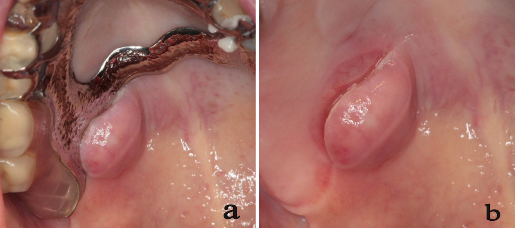
Figure 1. Intra-oral view. Clinical views of the lesion with (a) and without (b) the removable prosthesis. These views show a nodulary sessile mass on soft palate with inflammatory aspect on the surface and related to the prosthetic edge.
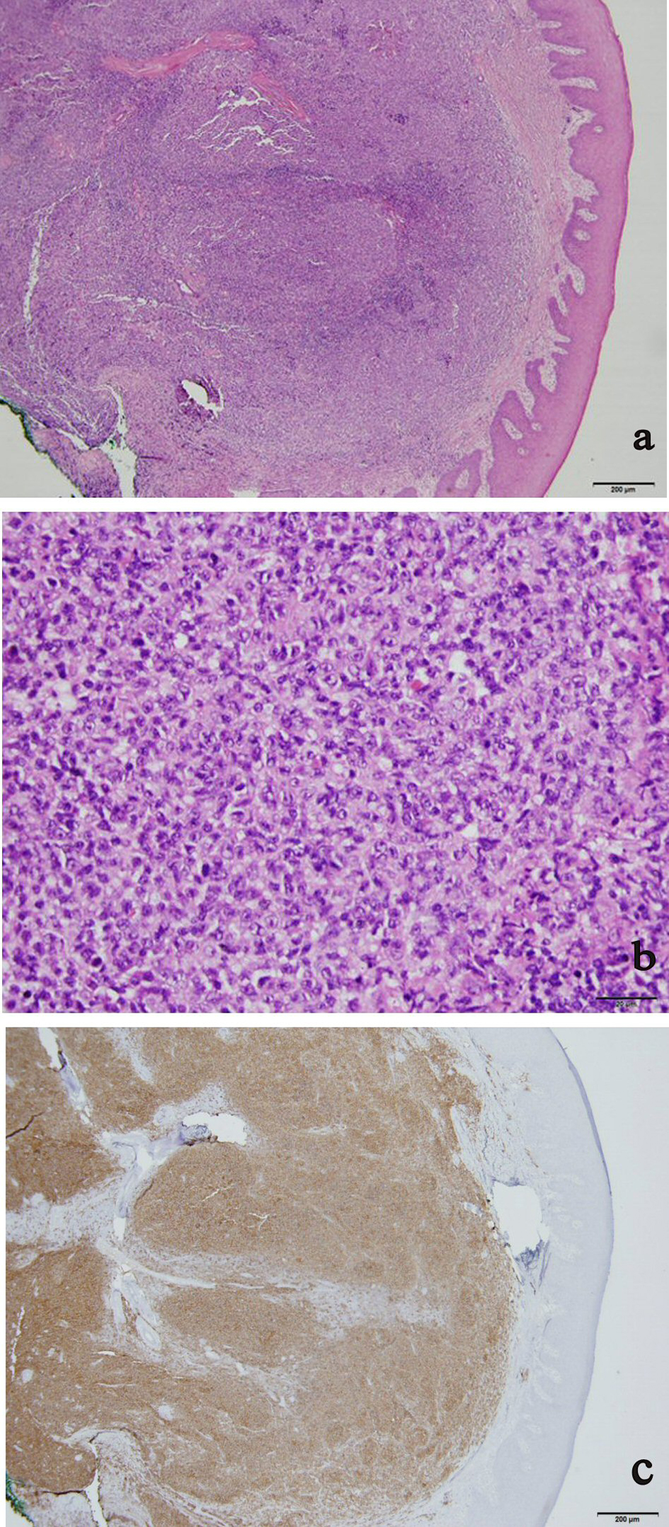
Figure 2. Microscopical views. Slides 2a (hematoxylin-eosin staining (HE), × 4) and 2b (HE, × 40) showing proliferation of lymphoid cells of small sizes (centrocytes) and of 10 to 15 of big sizes cells (centroblast) by field of view organized in follicular architecture and without tangible body macrophages; oral mucosa is covered by a normal malpighian epithelium; slide 2c (immunochemistry, × 4) shows a strong positivity to CD20 antibody compatible with B cells infiltration.
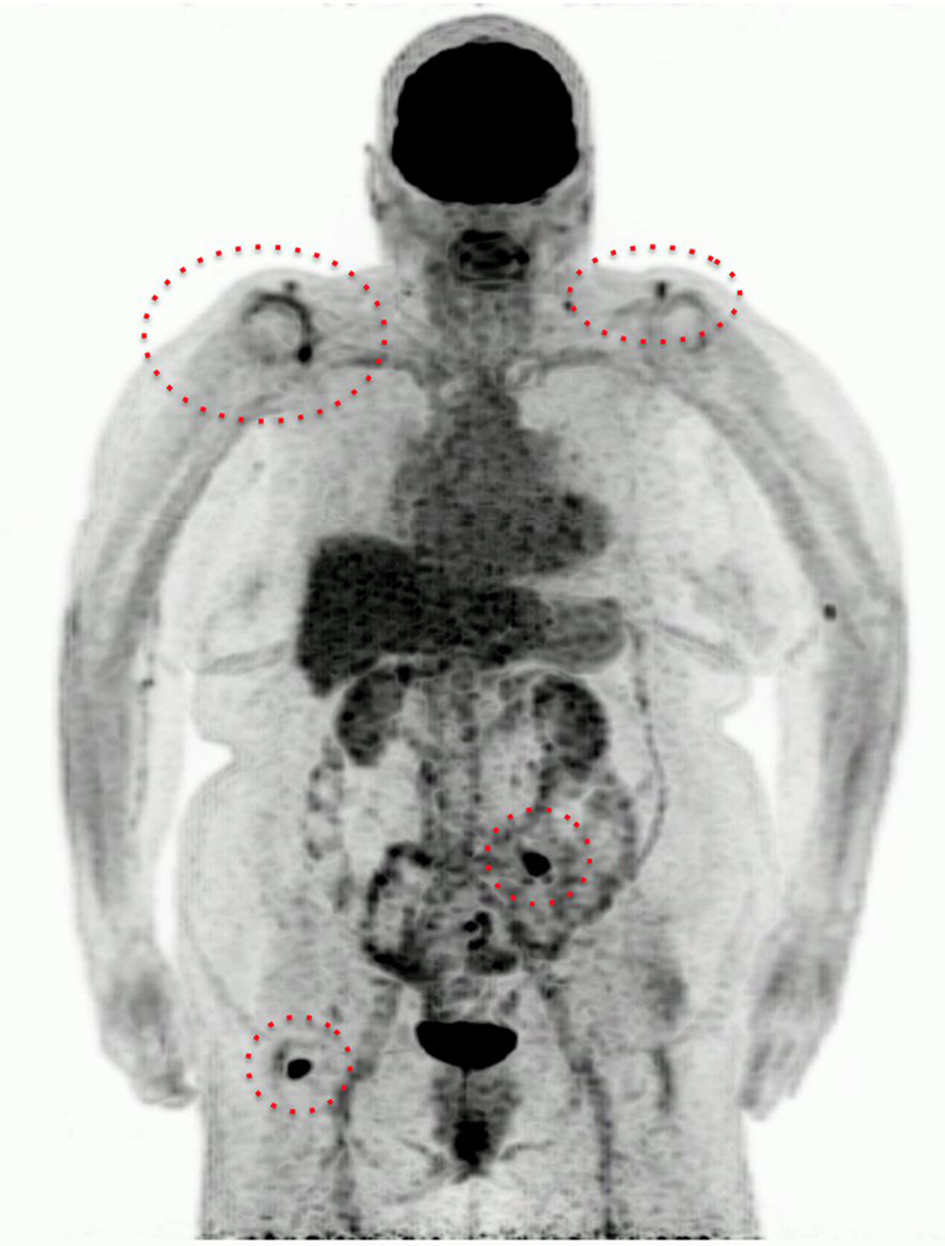
Figure 3. The 18-FDG PET-scanner of case 1. In cephalic region, we note a focal hyperfixation into the right palate which can correspond to a post-operative inflammation. In the thoraco-abdomino-pelvic region, there are two clavicular, one mesenteric and one right inguinal nodes.
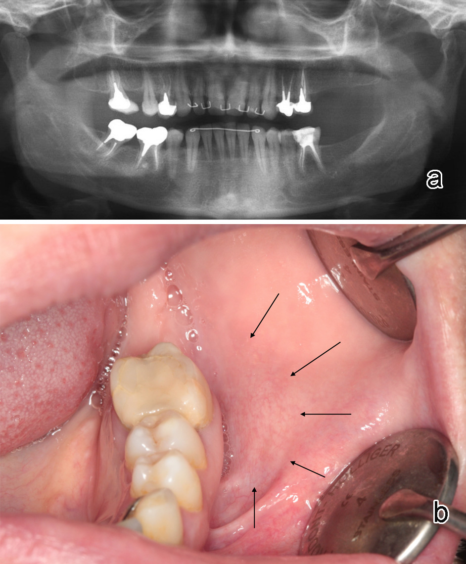
Figure 4. Clinical and radiological presentation of case 2. No dental infection in relation to buccal swelling (b) is observed on panoramic X-ray (a).
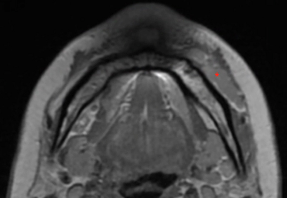
Figure 5. Facial magnetic resonance imaging (MRI) of case 2. Axial slice through mandibular body showing an elongated and well limited mass (*) slightly enhanced by gadolinium in T1 weighted image and laying in the buccal corridor against the buccal cortex.
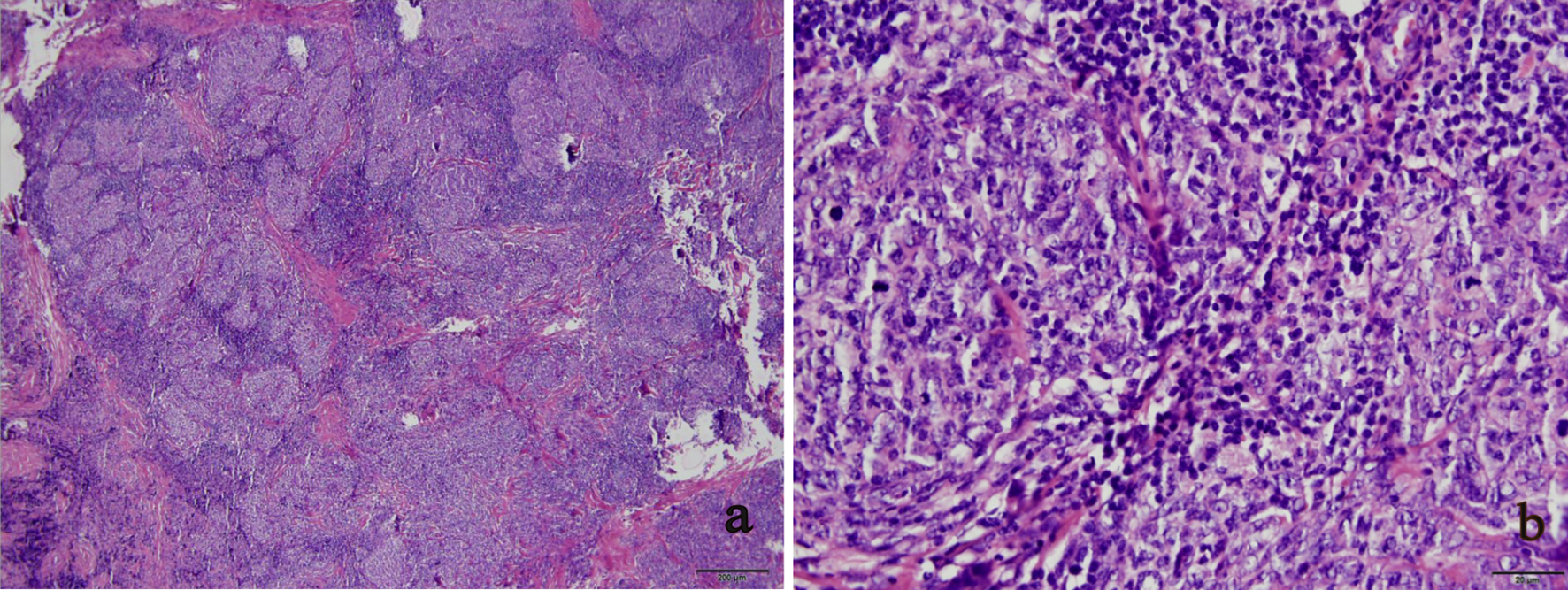
Figure 6. Microscopical views of case 2. (a) Lymphoid proliferation clustered into identifiable nodules (HE, × 4). (b) Centroblast cells are mainly observed in the field of view with many mitoses (HE, × 40).
Table
Table 1. Immunohistochemical Pattern
| Markers | Case1 | Case2 |
|---|
| CD: cluster designation. *Positivity to this three immunohistological markers features follicular lymphoma. |
| CD20* | ++ | + |
| bcl-6* | + | + |
| CD10* | + | +/- |
| bcl-2 | + | - |
| CD23 | + | + |
| CD5 | - | - |
| CD3 | - | - |
| Cycline D1 | - | / |
| Ki67 | 30% | 70% |