Journal of Hematology, ISSN 1927-1212 print, 1927-1220 online, Open Access
Article copyright, the authors; Journal compilation copyright, J Hematol and Elmer Press Inc
Journal website http://www.thejh.org

Case Report

Volume 5, Number 2, June 2016, pages 70-73


Pulmonary Thromboembolism Associated With Mixed-Type Autoimmune Hemolytic Anemia

Figures

Figure 1.
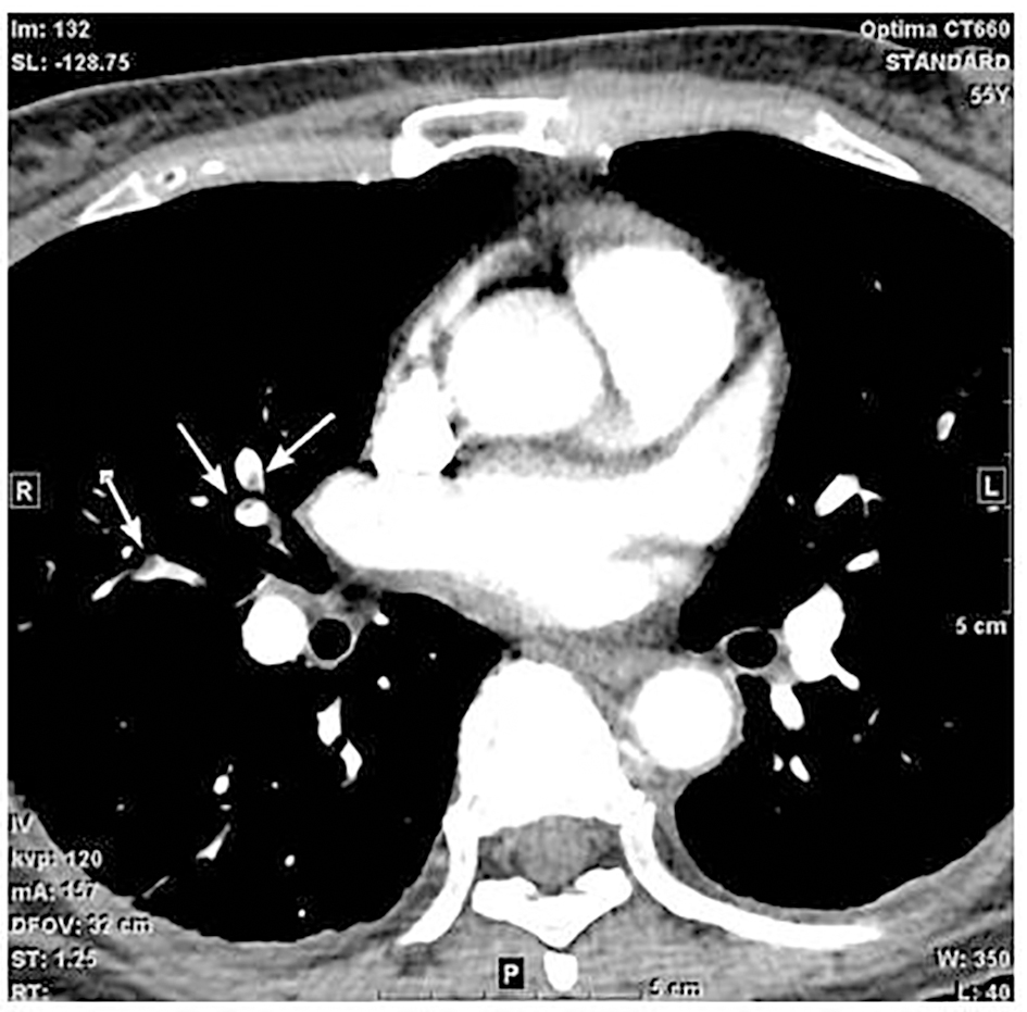
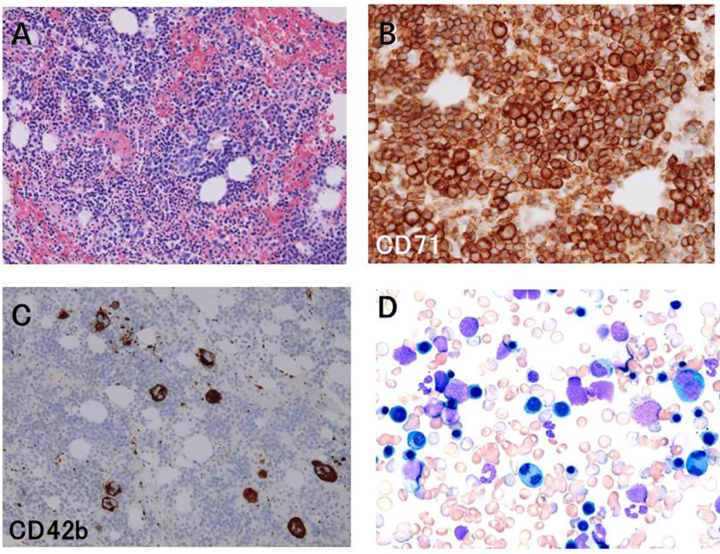
Figure 1. Examination of clot section specimen (A, B, and C) and wedge smear cytology (D) of the bone marrow aspirate. Hematoxylin and eosin stain (A) showed hypercellular bone marrow (× 100). Immunostaining with antibodies against CD71 (B) and CD42b (C) revealed that the eythroid cells were markedly increased and the megakaryocytes were also increased (× 200). Immature and binucleated erythroid cells were frequently observed (D, × 200).
Figure 2.
Figure 2. Contrast-enhanced computed tomography (CT) of the chest showed multiple filling defects in the bilateral pulmonary arteries (arrows). These findings are diagnostic of multiple acute pulmonary thromboembolism.

Table

Table 1. Thermal Amplitude of the Cold Agglutinin
 
Dilution× 1× 2× 4× 8× 16
The patient’s serum was serially diluted in normal saline and incubated with O type RBCs at room temperature, 4, 30, and 37 °C for 45 min and agglutination was determined. RT: room temperature.
4 °C2+2+1+Weak0
RT1+Weak000
30 °C1+1+000
37 °CWeak0000