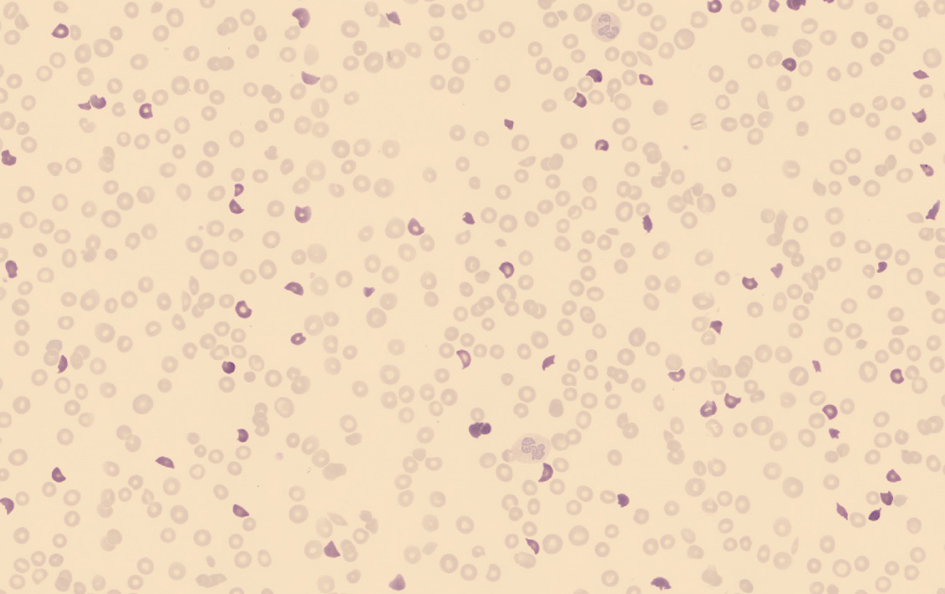
Figure 1. An overview of the red blood cells in a peripheral blood smear used for the classification in which the schistocytes (highlighted) are pre-classified by the software without manual intervention.
| Journal of Hematology, ISSN 1927-1212 print, 1927-1220 online, Open Access |
| Article copyright, the authors; Journal compilation copyright, J Hematol and Elmer Press Inc |
| Journal website http://www.thejh.org |
Letter to the Editor
Volume 4, Number 2, June 2015, pages 184-186
Automated Detection and Classification of Schistocytes by a Novel Red Blood Cell Module Using Digital Imaging/Microscopy
Figures
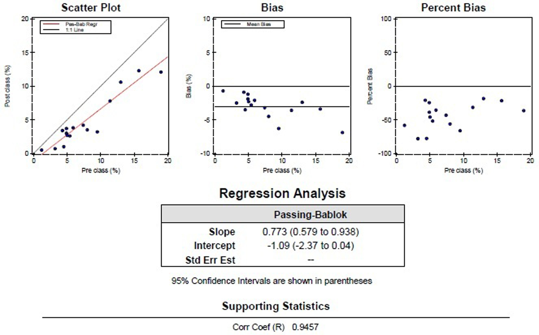
Table
| Diagnosis | Pre-classification in % | Post-classification in % |
|---|---|---|
| TTP | 13.0 | 10.6 |
| Solid tumor | 4.9 | 3.7 |
| 5.0 | 2.7 | |
| 15.7 | 12.3 | |
| 19.0 | 12.1 | |
| Thalassemia | 9.5 | 3.2 |
| 7.4 | 4.2 | |
| 11.4 | 7.8 | |
| Myelofibrosis | 5.9 | 3.8 |
| MDS-RAEB 2 | 4.5 | 1.0 |
| 4.9 | 3.0 | |
| CLL | 5.4 | 2.6 |
| Mantle cell lymphoma | 1.2 | 0.5 |
| Unknown | 3.2 | 0.7 |
| 8.0 | 3.5 | |
| 4.3 | 3.4 |