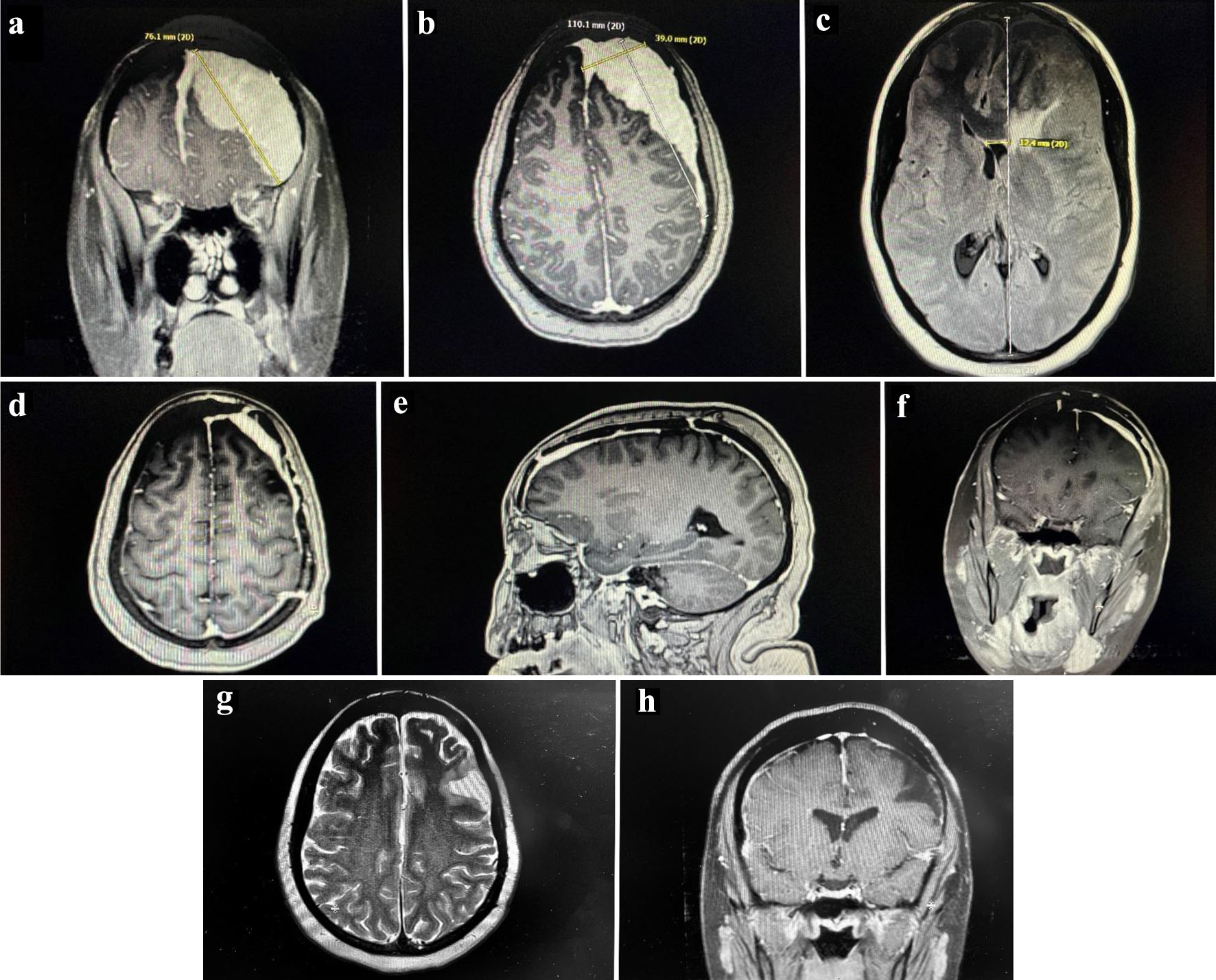
Figure 1. Pre-operative MRI scan. Dural-based homogeneously enhancing extra-axial mass along the left hemisphere with extension to the anterior interhemispheric falx and anterior right frontal lobe measuring 11 cm. There is significant mass effect and vasogenic edema. (a) Coronal section of the mass. (b) Sagittal section of the mass. (c) Transverse section of the mass. Post-operative MRI scan. Diffuse dural thickening through the left hemisphere, most notably along the left frontal lobe, reflecting postoperative changes of residual tumor. (d) Transverse section. (e) Sagittal section. (f) Coronal section. Post-chemotherapy MRI scan. Unchanged dural thickening and enhancement along the left hemisphere and anterior interhemispheric falx post-operatively. Some encephalomalacia in the left frontal lobe, but no new pathological enhancement seen. (g) T2 transverse section. (h) T1 axial section. MRI: magnetic resonance imaging.