Journal of Hematology, ISSN 1927-1212 print, 1927-1220 online, Open Access
Article copyright, the authors; Journal compilation copyright, J Hematol and Elmer Press Inc
Journal website http://www.thejh.org

Case Report

Volume 7, Number 1, January 2018, pages 38-42


Mantle Cell Lymphoma Relapsing as Disease of Skin, Orbit and CNS: An Extremely Rare Presentation and a Review of Literature

Figures

Figure 1.
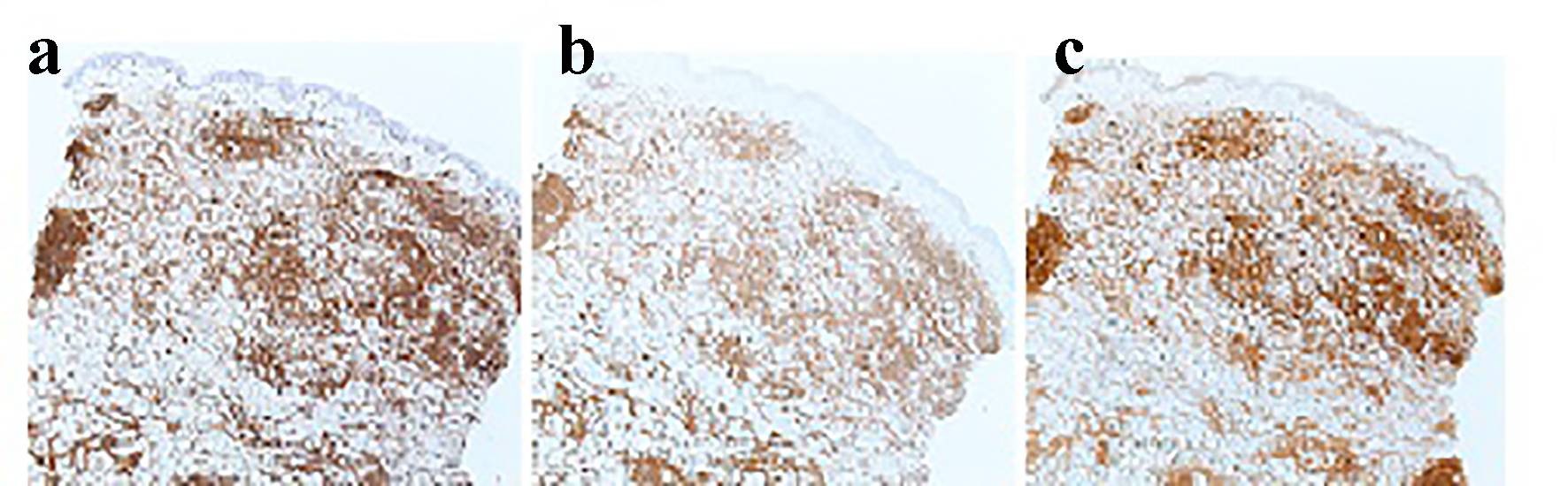
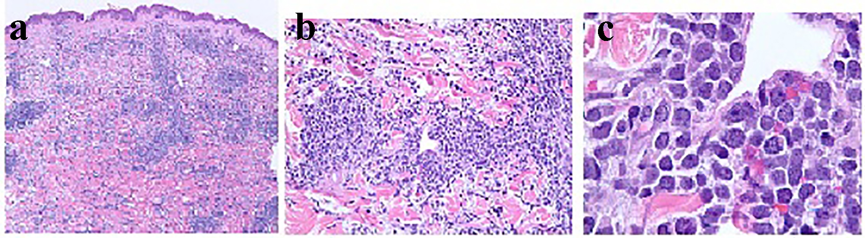
Figure 1. Histopathology of the skin on biopsy with × 4, × 20 and × 100 magnifications in (a), (b) and (c), respectively, demonstrating dense infiltrate of atypical lymphoid cells distributed throughout the dermis and subcutis. The atypical lymphocytes appear as small to medium sized, contain scant cytoplasm, and have hyperchromatic nuclei with irregular nuclear contours and occasional indistinct nucleoli. There are scattered apoptotic bodies and occasional mitotic figures.
Figure 2.
Figure 2. Immunostain of skin biopsy: (a) expression of CD5 by neoplastic cells (× 4); (b) expression of CD20 (× 4); (c) expression of cyclin D1 by the malignant lymphoid cells (× 4).
Figure 3.
Figure 3. CSF cytology with × 10 and × 100 magnifications in (a) and (b), respectively, showing cells suspicious for lymphoma which stained positive for cyclin D1 immunostain.