Journal of Hematology, ISSN 1927-1212 print, 1927-1220 online, Open Access
Article copyright, the authors; Journal compilation copyright, J Hematol and Elmer Press Inc
Journal website http://www.thejh.org

Case Report

Volume 6, Number 4, October 2017, pages 101-104


Revisiting Howell-Jolly Body-Like Cytoplasmic Inclusions in Neutrophils: A Report of Two Cases and Confirmation of Nuclear Origin

Figures

Figure 1.
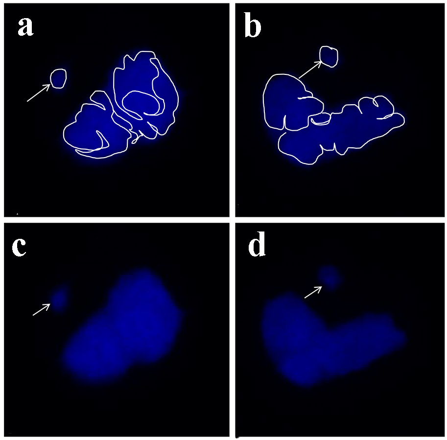
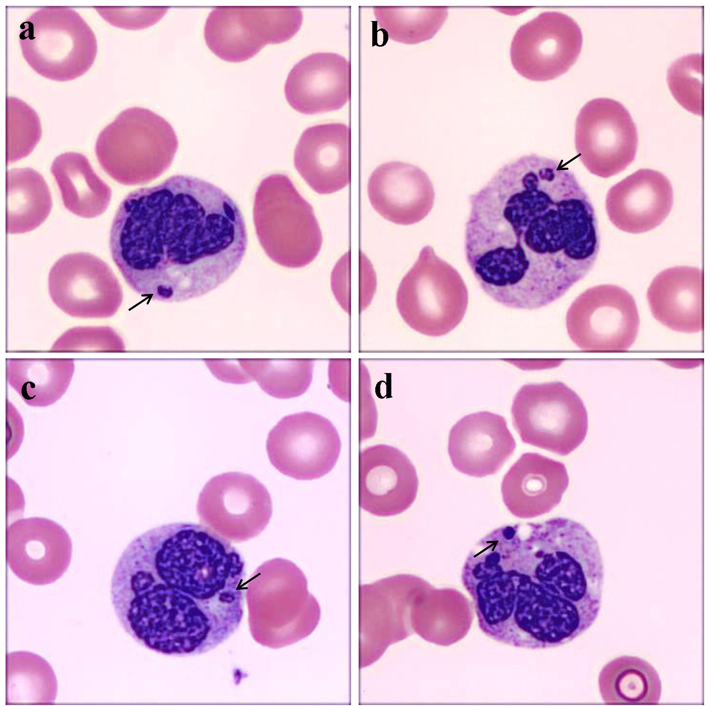
Figure 1. (a-d) Spherical Howell-Jolly body-like inclusions within neutrophil cytoplasm from case 1 (a and b) and 2 (c and d) highlighted by short black arrows. These inclusions are separate from the main nuclear body and demonstrate a similar basophilic staining pattern on the Wright Giemsa stain. The main nucleus demonstrates a coarsely condensed chromatin. The secondary neutrophilic granules demonstrate some toxic change, more pronounced in d.
Figure 2.
Figure 2. 4′,6-diamidino-2-phenylindole (DAPI) immunofluorescence microscopy images demonstrating a single distinct spherical nuclear fragment consistent with the Howell-Jolly body-like inclusion (arrows) located separate from the neutrophil nucleus in two representative nuclei in two representative neutrophils (panels a and b with nuclear features highlighted, and c and d demonstrate the same two neutrophils without highlights).