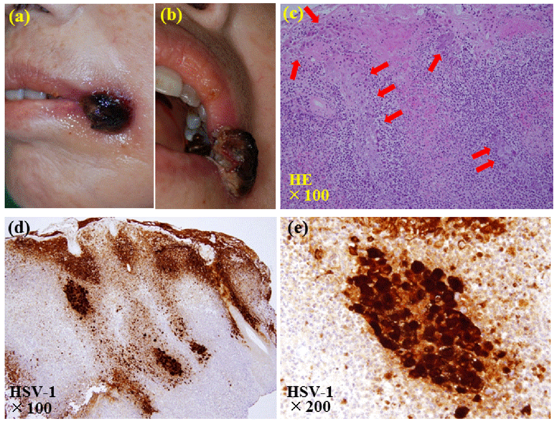
Figure 1. Pictures and pathological findings of the nodule at the left angle of the mouth. (a, b) A painful nodule of 1.5 cm in size with black necrotic tissue is seen at the left angle of the mouth. (c) The biopsy of the nodule reveals ulcerative lesions with necrosis of the epithelium and vesicular formation. Enlarged epithelial cells whose nuclei are enlarged with moldering and displacement of chromatin to the periphery, and intranuclear inclusion bodies are seen (arrows) (hematoxylin and eosin stain, × 100). (d, e) Immunostaining shows that these epithelial cells are positive with antibody against anti-herpes simplex virus type 1 (d: × 100; e: × 200).