Figures
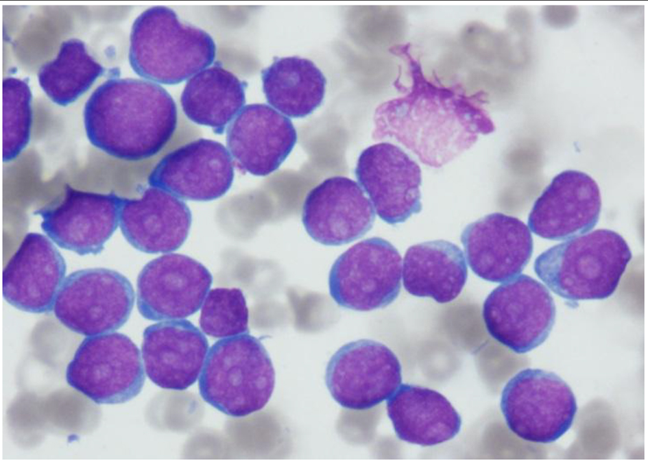
Figure 1. Aspirate smear showing extensive infiltration with blasts, small in size with very high nucleocytoplasmic ratio and multiple prominent nucleoli. Few blasts show fine cytoplasmic blebs/pseudopods (Wright stain, × 1,000 magnification).
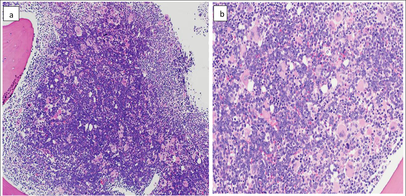
Figure 2. Bone marrow biopsy (H&E) showing marked hypercellularity (about 100%) and diffuse infiltration with monomorphic population of small to medium sized lymphoid blasts. The blasts in the center of the biopsy appear larger with more prominent nucleoli compared to periphery (a) (× 10). Marked megakaryocytic hyperplasia (b) (× 20).
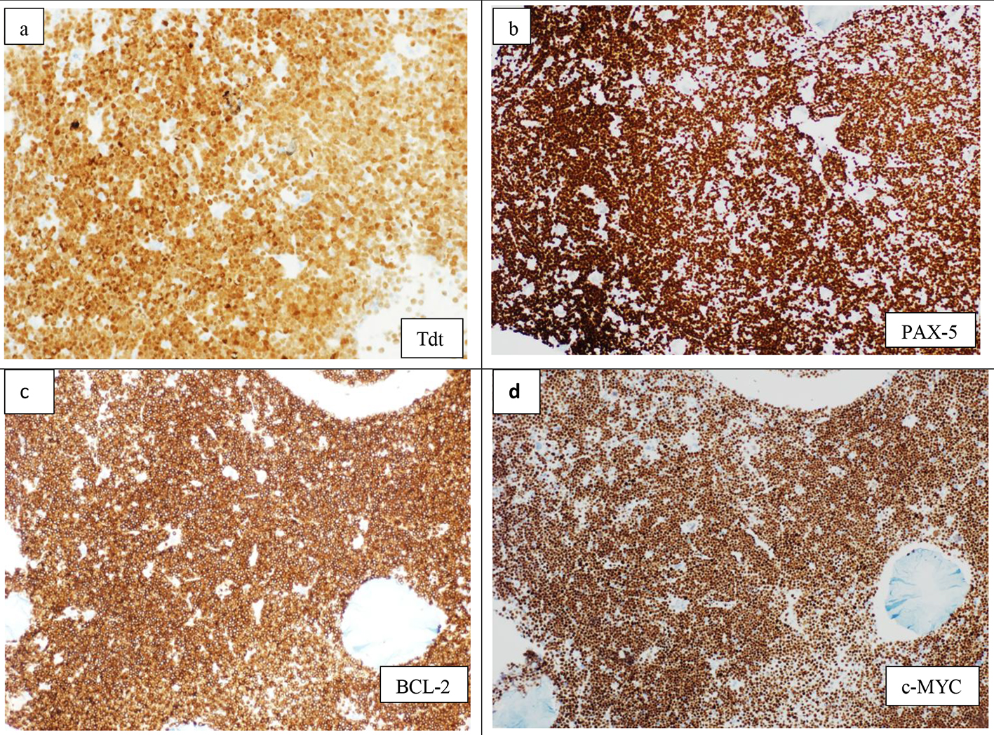
Figure 3. Immunohistochemistry on bone marrow biopsy showing diffuse infiltration by lymphoid blasts, positive for TdT (× 20) (a), PAX-5 (× 10) (b), BCL-2 (×10) (c), and c-MYC (× 10) (d).
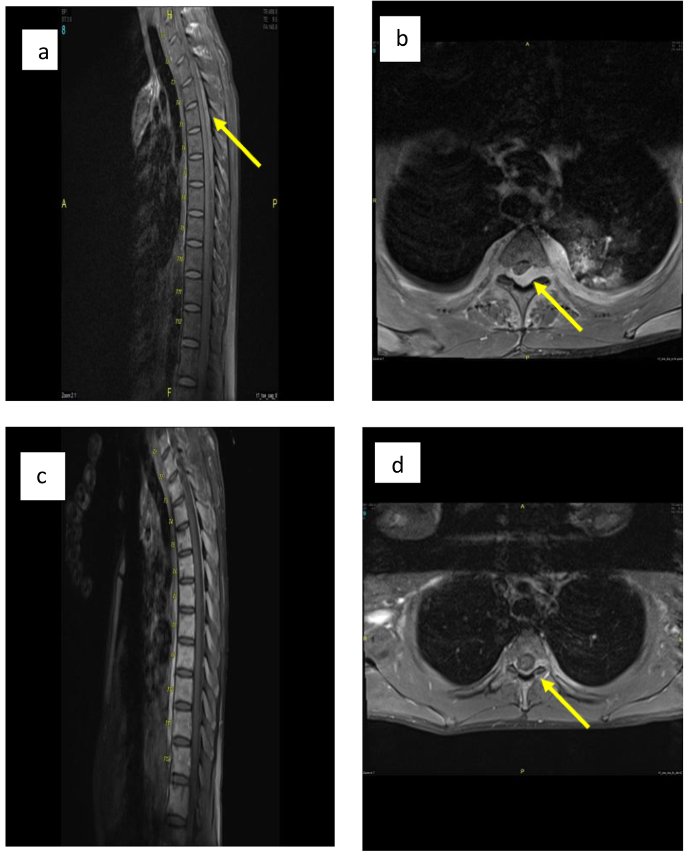
Figure 4. (a, b) Sagittal and axial post-contrast fat saturated images of thoracolumbar spine before treatment show posterior intraspinal extradural mass lesion extending from T2 to T8 levels in sagital image. Mildly compression are seen at the posterior aspect of cord, more from left side at T3 in axial image (arrow in b) with extension into both neural foramina more in left side as well as small paraspinal soft tissue component. (c, d) Sagittal and axial post-contrast fat saturated images at the same level after treatment show significant improvement with only small residual component seen posterior to cord at T3 level and extending into left neural foramina (arrow in d).