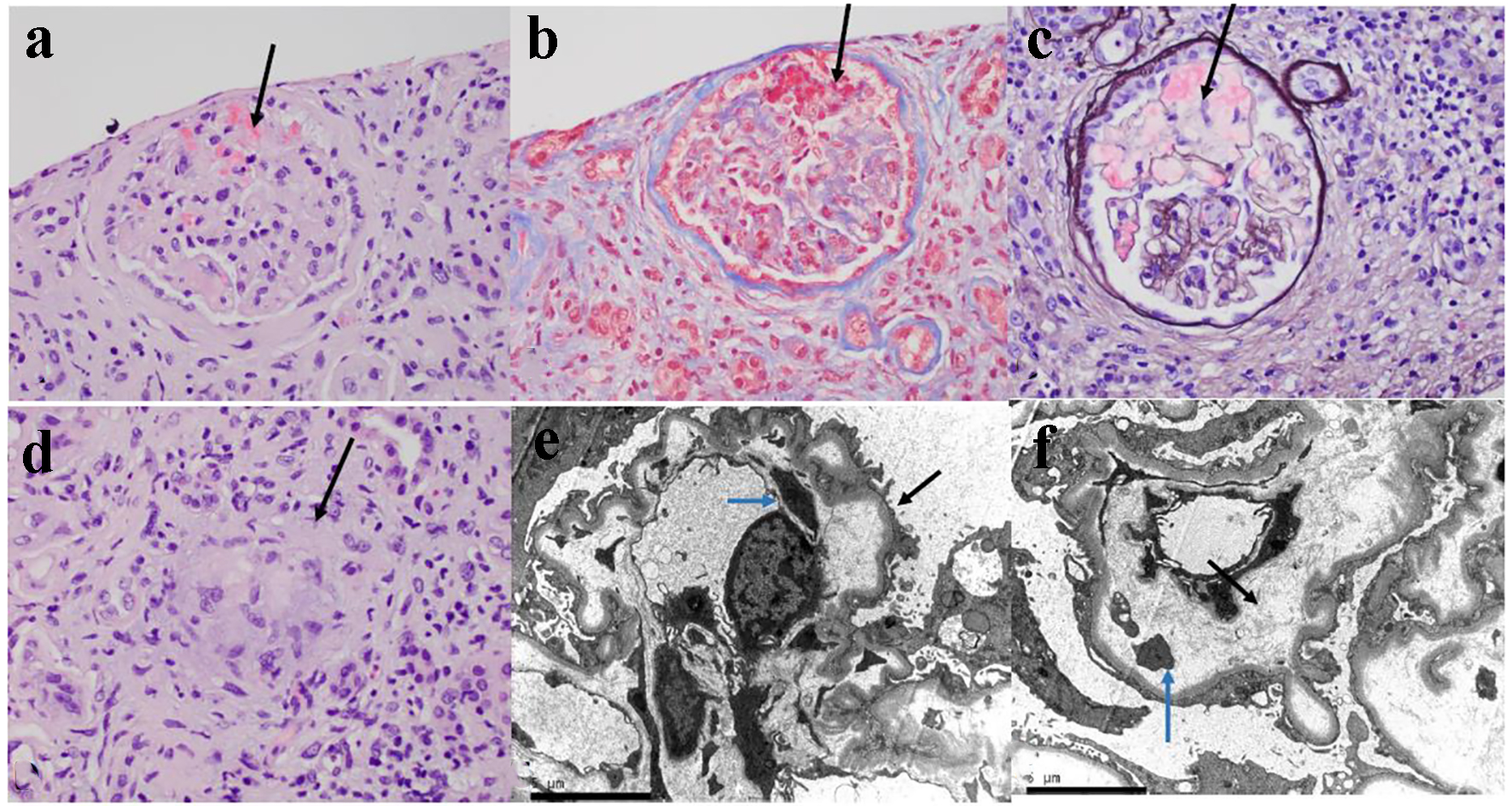
Figure 3. The kidney biopsy tissue of the patient. (a) The glomerulus is slightly hypocellular, and most of the glomerular capillary lumina are closed due to thickening of the capillary walls. Red blood cells and fragmented cells are seen in the mesangial area (H&E, × 400). (b) Trichrome stain showing microthrombi in glomerular capillaries and focal reduplication of the glomerular capillary basement membranes (× 400). (c) Ectatic glomerular capillary lumina are present as a result of mesangiolysis. Focal reduplication of the glomerular capillary basement membranes is also present (Jones silver stain, × 400). (d) Focal non-caseating granuloma is present in the interstitium (black arrow, H&E, × 400). (e) Electron microscopy of glomerular capillary loops showing podocytes vacuolization, extensive effacement of foot processes (black arrow) and basement membrane reduplication (blue arrow). (f) Electron microscopy of glomerular capillary loops showing markedly narrowed capillary lumen and widening of the subendothelial spaces with electrolucent fluffy material (black arrow) and platelets (blue arrow).