Journal of Hematology, ISSN 1927-1212 print, 1927-1220 online, Open Access
Article copyright, the authors; Journal compilation copyright, J Hematol and Elmer Press Inc
Journal website http://www.thejh.org

Case Report

Volume 4, Number 4, December 2015, pages 238-241


Diffuse Intrasinusoidal Hepatic Metastasis From Occult Breast Carcinoma Presenting as Thrombotic Microangiopathy: A Case Report and Literature Review

Figures

Figure 1.
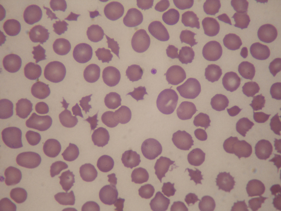
Figure 1. Peripheral blood film showing numerous keratocytes, schistocytes and polychromasia, in keeping with microangiopathic hemolysis.
Figure 2.
Figure 2. MRI of the liver. (a) T1 with fat saturation, (b) T2, (c) T1 + contrast with fat saturation, (d) apparent diffusion co-efficient (ADC). Representative images show T1 hypo-intense (thin arrows) and T2 hyper-intense (dotted arrows) ill-defined lesions distributed in a fairly linear fashion in the right hepatic lobe. The lesions do not show significant contrast enhancement (thick arrows) or restricted diffusion (black arrows).
Figure 3.
Figure 3. DISH. High power magnification showing well-defined rounded nests of intrasinusoidal tumor cells with enlarged nuclei, moderately abundant cytoplasm and an occasional mitotic figure (arrow). The adjacent hepatic parenchyma on the left shows atrophy of the liver cords indicative of ischemic change (hematoxylin and eosin, × 400). Inset: immunohistochemical staining with AE1/3, a cytokeratin stain, confirming the epithelial nature of the intrasinusoidal malignant cells (AE1/3 stain, × 200).