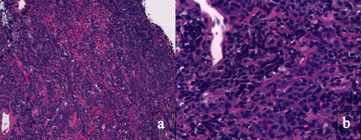
Figure 1. Histology of tissue biopsy of duodenum. Photomicrographs show the tumor in the duodenum under (a) low magnification × 4 and (b) high magnification × 20 on hematoxylin and eosin staining. PBL is characterized by monomorphic cellular proliferation of round or oval cells with centrally or eccentrically placed nuclei and abundant eosinophilic cytoplasm in a diffuse growth pattern. Apoptotic bodies and mitotic figures and macrophages can lead to a starry sky appearance.
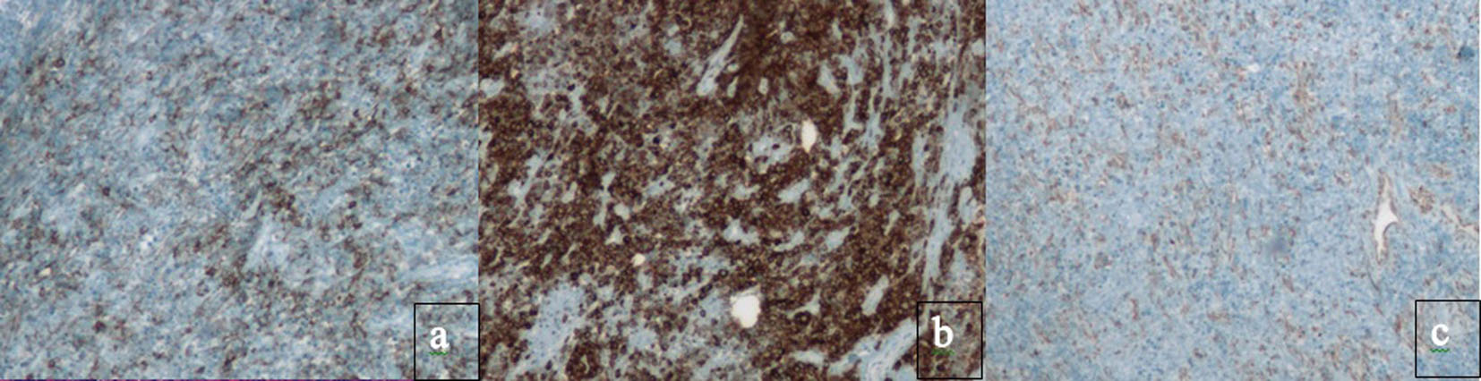
Figure 2. Immunohistochemical staining absent in PBL. Classic B cell markers, such as CD20, CD79a, PAX-5 and CD45, are lost. Immunohistochemical staining for (a) CD20, (b) CD79a and (c) PAX-5. Instead, PBL has the immunophenotype of a terminally differentiated B cell.
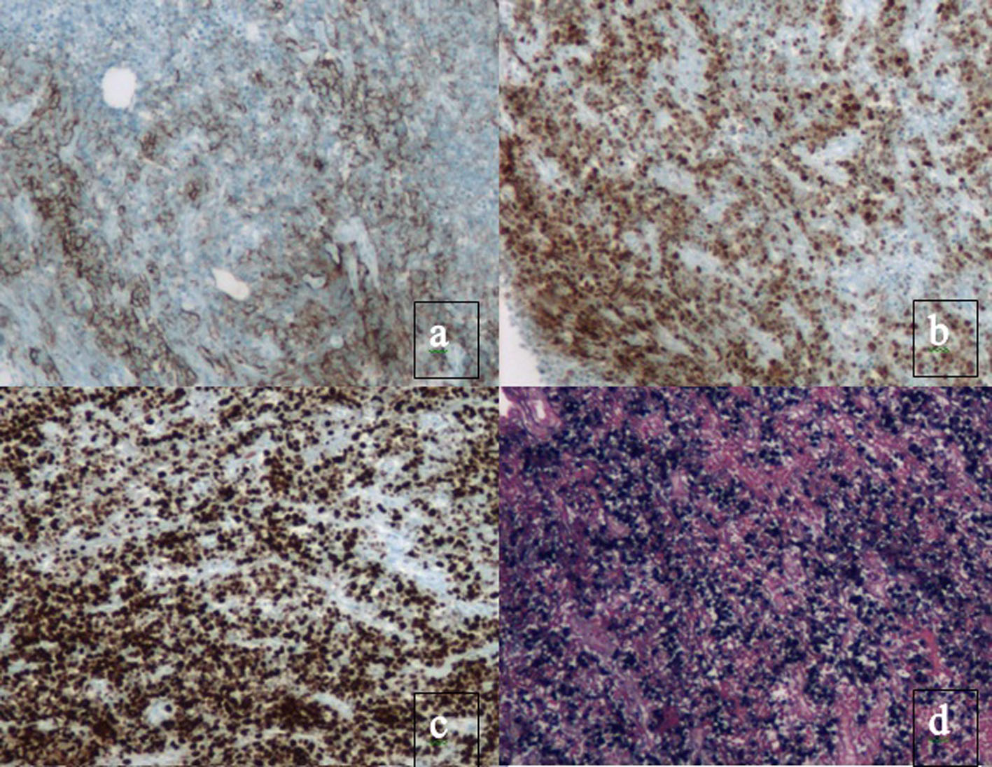
Figure 3. Immunohistochemical staining present in PBL. Plasma cell markers, such as CD38, CD138 and myeloma oncogene-1 (MUM1/IRF4) are expressed. There is also a high proliferation rate reflected by Ki67 expression > 80% and EBV present. Immunohistochemical staining for (a) CD138, (b) MUM1, (c) Ki67 and (d) EBER.