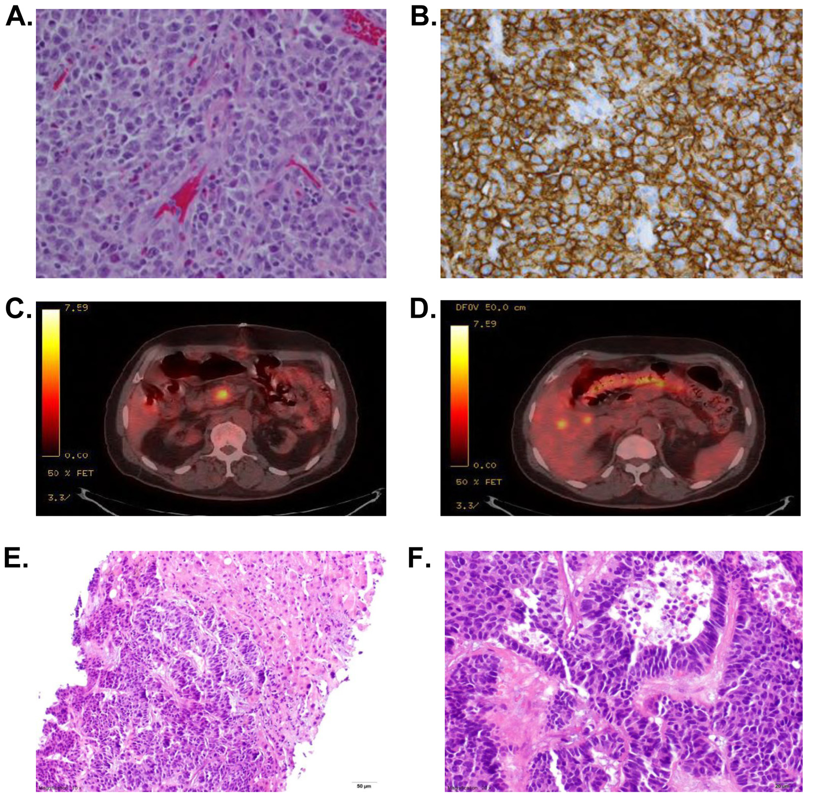
Figure 1. (A, B) Diffuse large B cell lymphoma. (A) Hematoxylin and eosin (H&E) stain at × 40 of resected tumor from terminal ileum showing sheets of large, pleomorphic cells with prominent nucleoli. (B) CD20 stain which was highly positive confirming this tumor to be of B cell origin. (C, D) Representative frames of the PET-CT demonstrating FDG uptake in liver and pancreatic head after resection of small bowel tumor. (E, F): PNET. Representative H&E stains from biopsy of a liver tumor from our patient demonstrating a nested and trabecular arrangement of neoplastic cells with high N:C ratios, frequent mitoses, focal necrosis and uniform chromatin at × 10 (E) and × 20 (F).